Discover more than 102 x ray heel lateral view latest
Details images of x ray heel lateral view by website dienmayquynhon.com.vn compilation. Orthobullets on Instagram: “Can you answer our FREE Question of the Day? A 17-year-old female presents with foot pain associated with footwear. A current lateral radiograph of the affected foot is depicted. CoLink Cfx Z-Plasty™. Cureus | A Review of Pediatric Heel Pain | Article. Ankle (horizontal beam lateral view) | Radiology Reference Article | Radiopaedia.org
 Fractures and dislocations of the tarsal bones | Anesthesia Key – #1
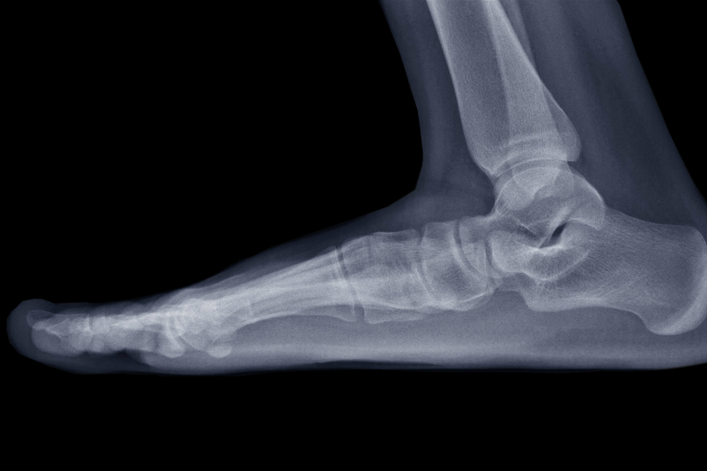 Plantar Heel Pain Workup: Laboratory Studies, Imaging Studies – #2
 Osgood–Schlatter disease – Wikipedia – #3
 196 Foot Film Photos, Pictures And Background Images For Free Download – Pngtree – #4
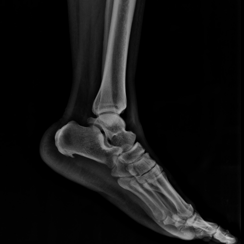 Ankle x-rays – Don’t Forget the Bubbles – #5
- x ray heel lateral view positioning
- calcaneus x ray anatomy
- normal heel x ray
 Interesting Cases – #6
 1,157 X Ray Calcaneus Images, Stock Photos, 3D objects, & Vectors | Shutterstock – #7
- lateral calcaneus positioning
- calcaneus lateral view position
- x ray heel ap view positioning
 Diagnostic Imaging Techniques of the Foot and Ankle | Musculoskeletal Key – #8
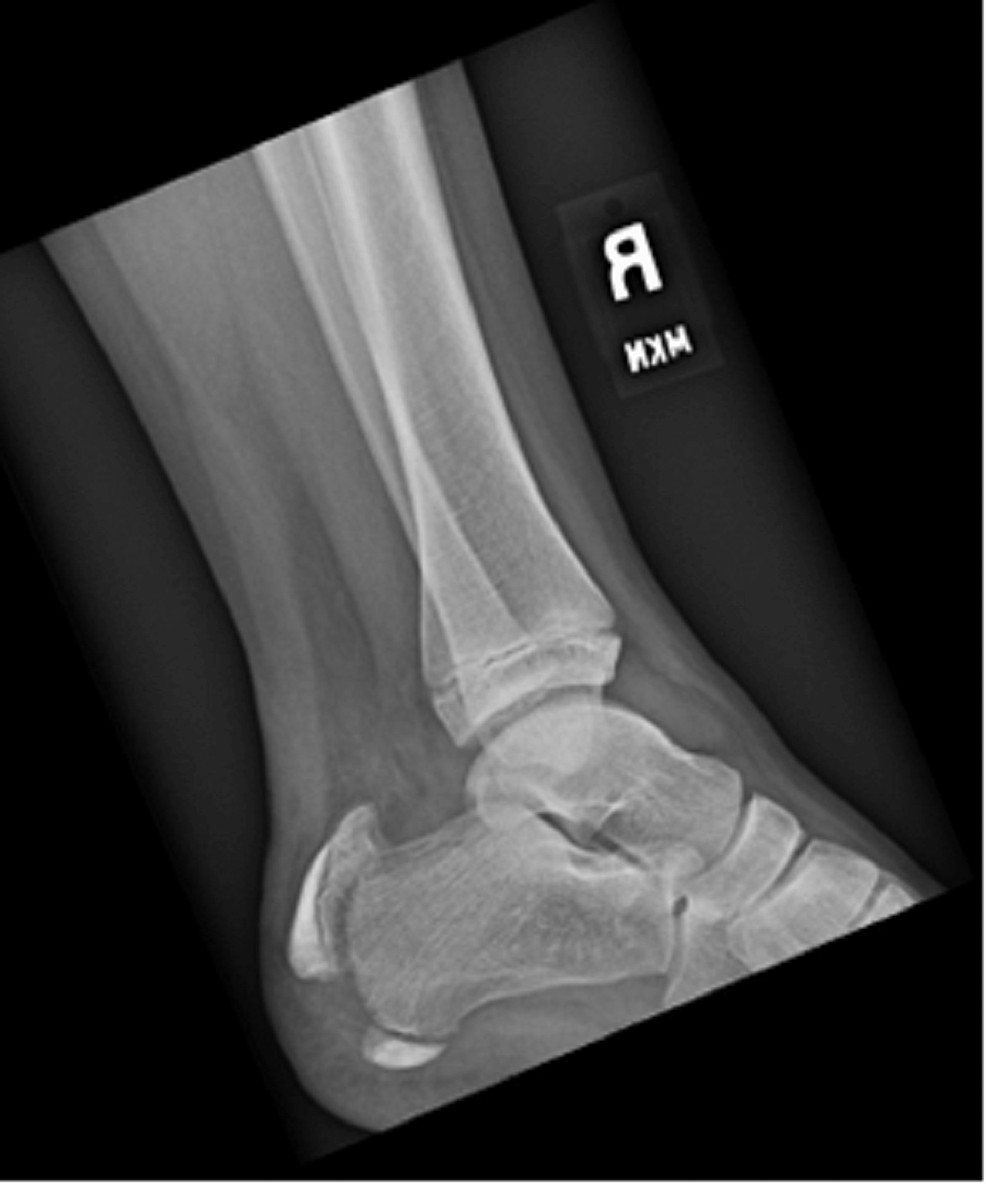 Congenital and Developmental Disorders of the Foot and Ankle | SpringerLink – #9
 Plantar Fasciitis and Heel Spurs: Understanding the Connection — Dr. Elton – #10
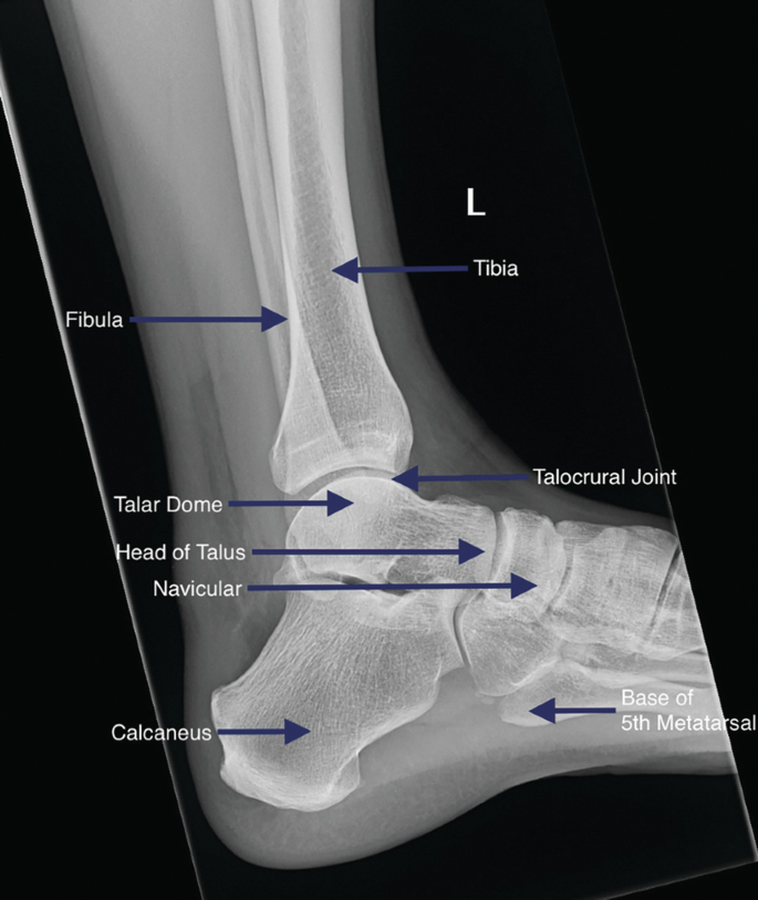 1,587 Xray Foot Stock Photos – Free & Royalty-Free Stock Photos from Dreamstime – #11
 Stress Fractures of the Calcaneus (Heel Bone) (Dr. Ed Davis, San Antonio Podiatrist – Heel Pain Blog) – #12
 Xray Image Ankle Lateral View Show Stock Photo 272065442 | Shutterstock – #13
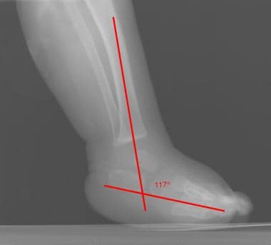 Anterior process calcaneal bone fracture | Radiology Case | Radiopaedia.org – #14
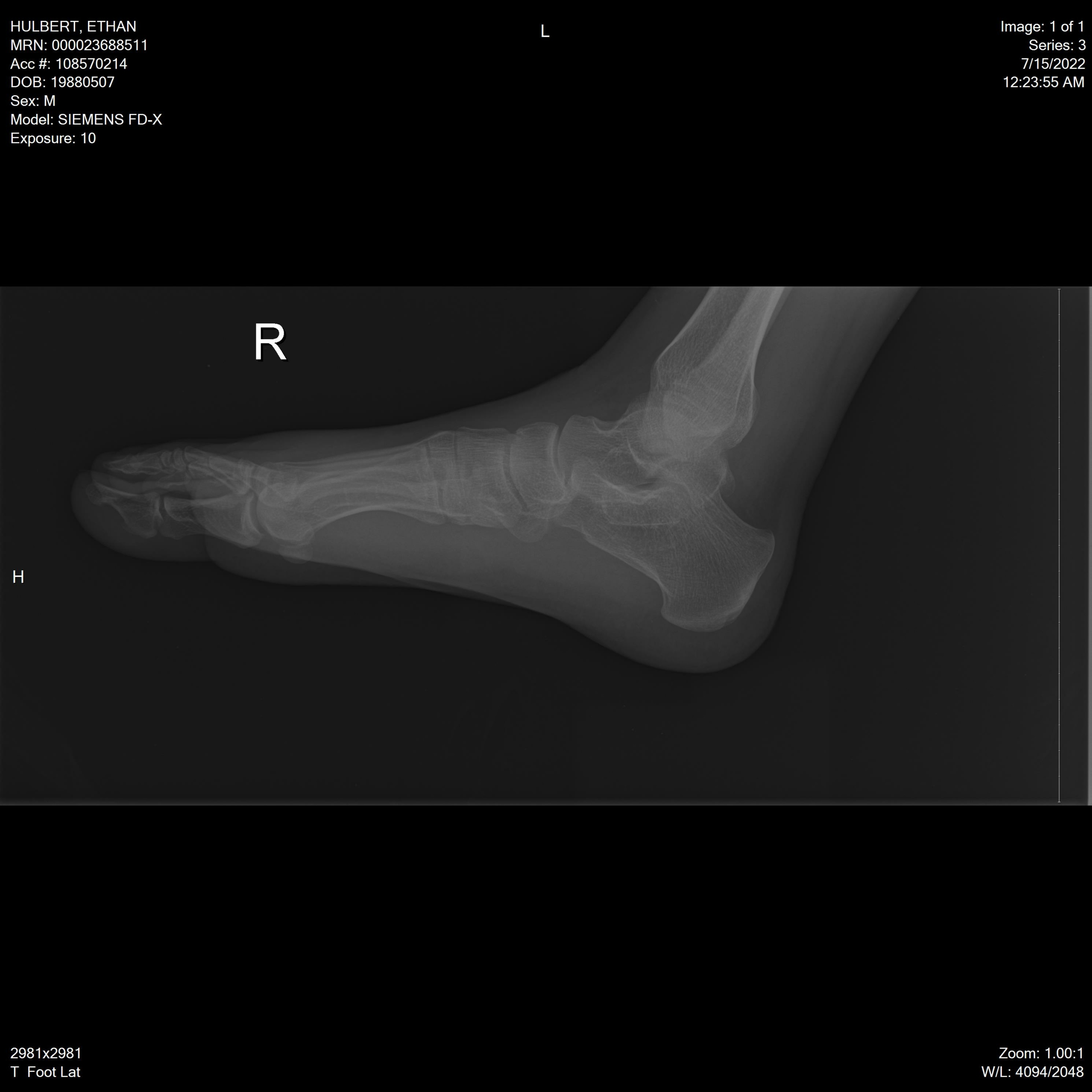 Calcaneal Fractures and Böhler’s Angle – JETem – #15
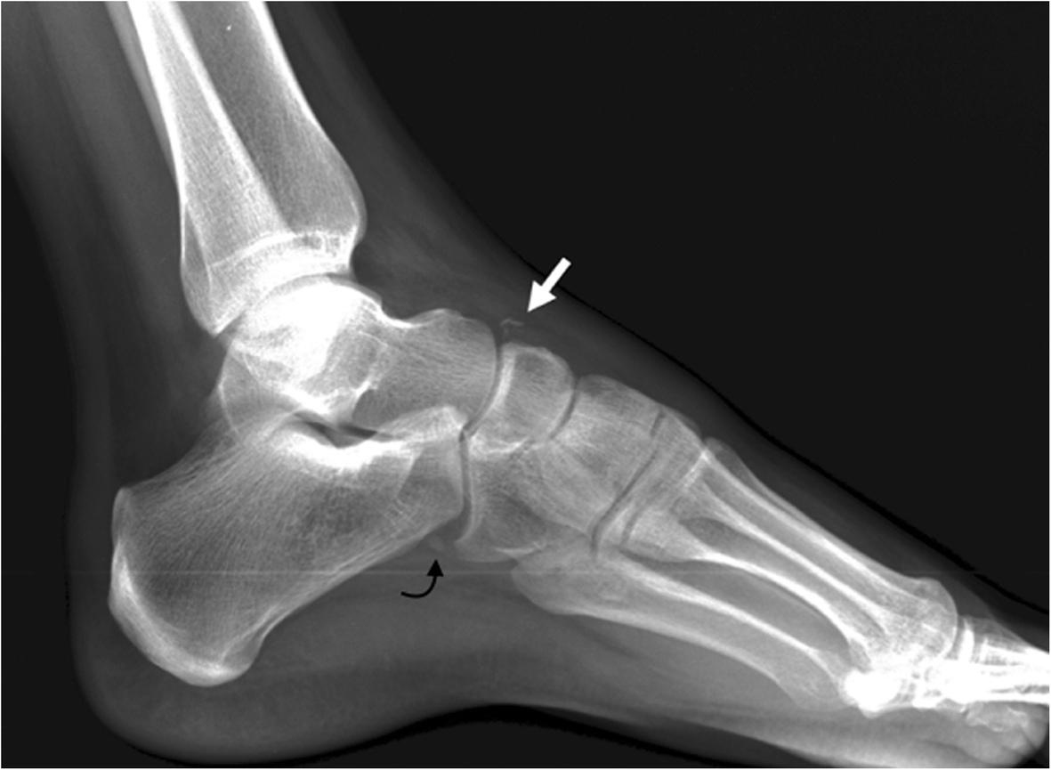 Xray Image Of Ankle Ap And Lateral View Stock Photo – Download Image Now – Metatarsus, Pain, 2015 – iStock – #16
 Insoles for unusual feet – Podiatry Clinics (Yorkshire) Ltd – #17
 BoneView – Gleamer – #18
- x ray left foot lateral view
- radiografia de pie roto
- normal heel x ray vs heel spur
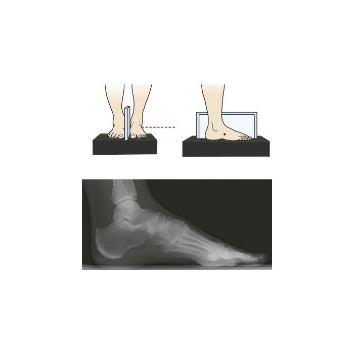 Radiology Quiz 44539 | Radiopaedia.org – #19
 Xray Image Broken Calcaneusheel Lateral View Stock Photo 269956949 | Shutterstock – #20
 TPLO Radiograph Positioning — Anchor Veterinary Surgery – #21
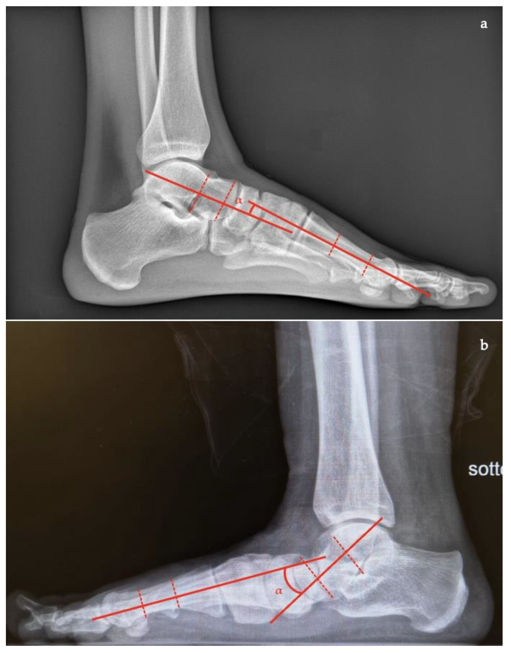
 Diagnostics | Free Full-Text | Adult Acquired Flatfoot Deformity: A Narrative Review about Imaging Findings – #22
- x ray foot ap lat
- calcaneal view position
- calcaneus lateral
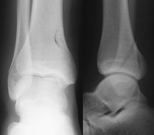 CALCANEUS LATERAL POSITIONING HINDI | X RAY POSITIONING FOR RADIOGRAPHERS | DOCTOR INSIDE – YouTube – #23
 The diagnostic dilemma of congenital foot deformity in pediatrics: could adding ultrasound be problem solving? | Egyptian Journal of Radiology and Nuclear Medicine | Full Text – #24
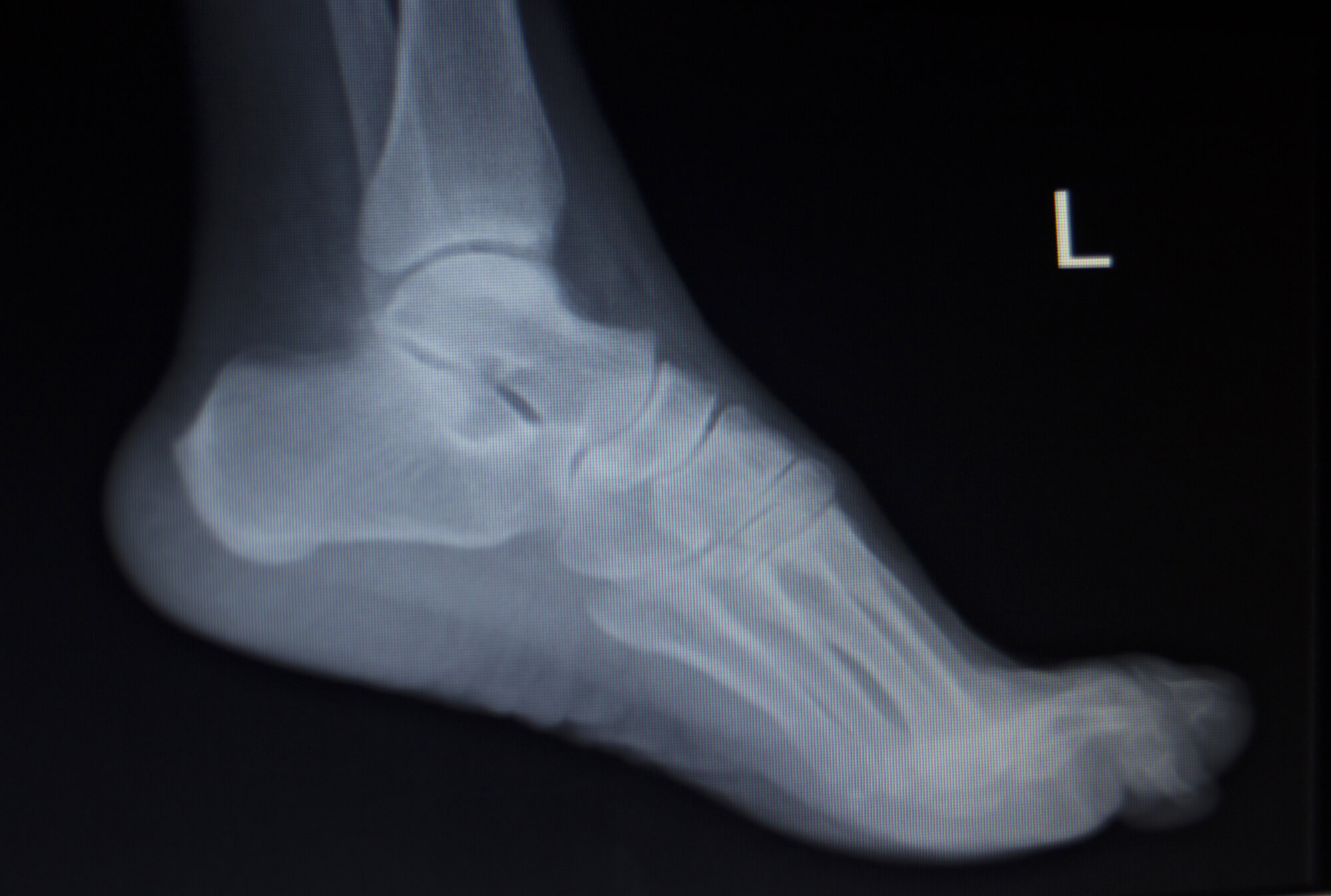
 Foot pain foot injury Xray of foot. Radi… | Stock Video | Pond5 – #25
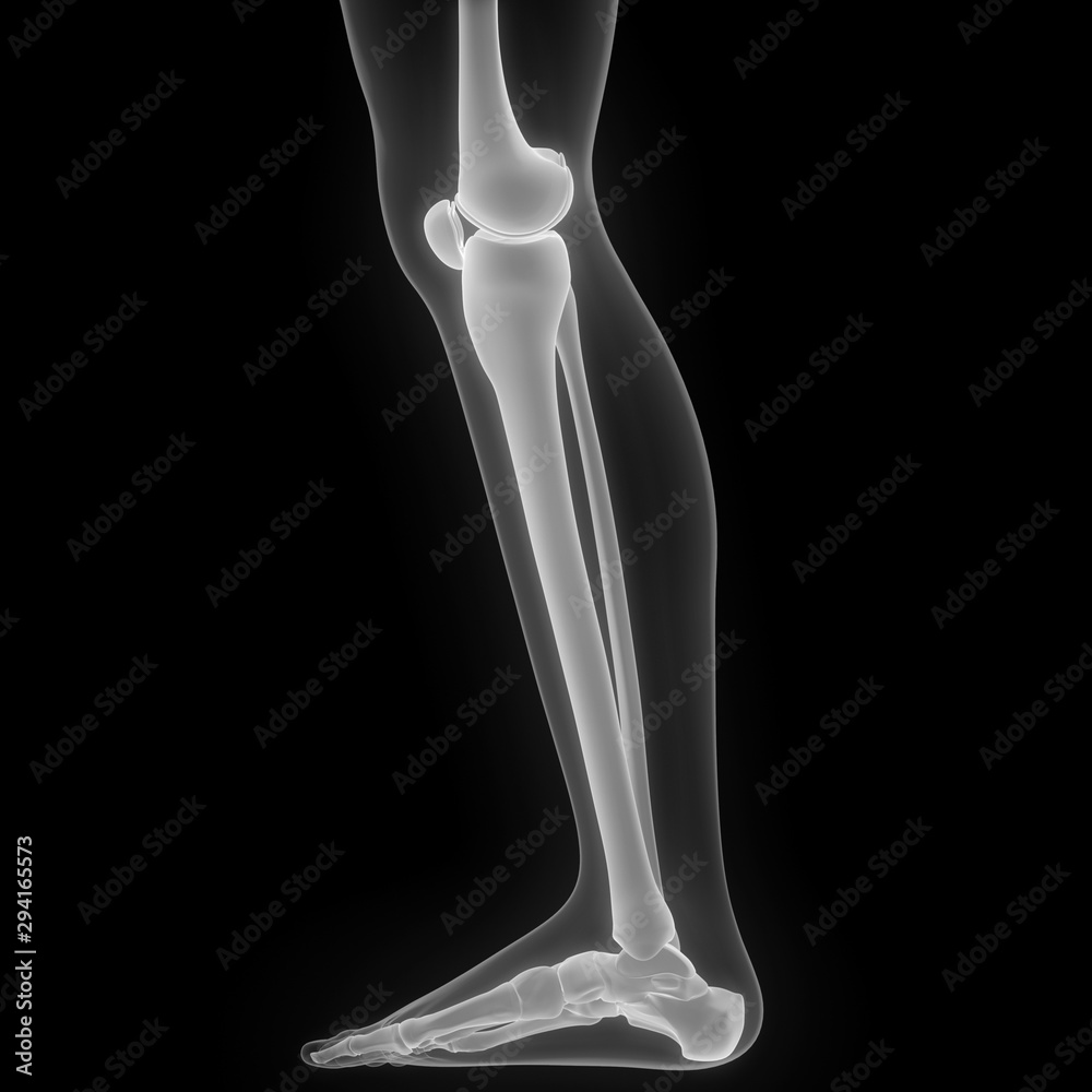 X-ray normal human’s foot lateral Stock Photo by ©stockdevil_666 61372863 – #26
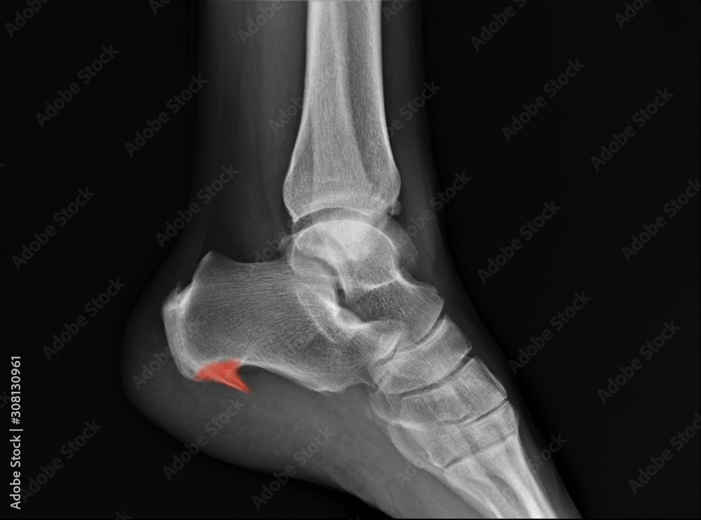
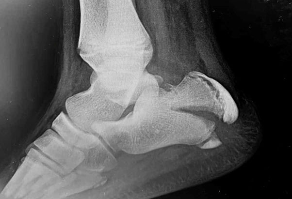 Film X-ray left lateral ankle radiograph showing spur at heel bone ( calcaneus). The calcaneal spur cause plantar fasciitis, heel pain (highlight on pain area). medical concept Stock Photo | Adobe Stock – #27
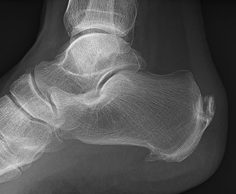 Lateral Foot Xray Image Stock Photo – Download Image Now – Spur, Calcaneus, Foot – iStock – #28
 Orthobullets – A lateral radiograph of the ankle demonstrating sclerosis of the navicular with abnormality of the talonavicular articulation. | Facebook – #29
 X-ray image of left ankle and foot , lateral view. Showing heel fracture. Stock Photo | Adobe Stock – #30
 Cureus | A Review of Pediatric Heel Pain | Article – #31
- heel ap position
- x ray heel ap lateral view
- normal heel xray child
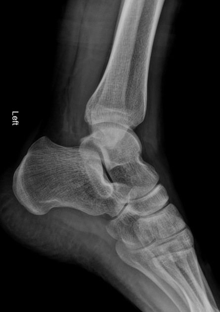 Radiographic measurements of the talus and calcaneus in the adult pes planus foot type – Agoada – 2020 – American Journal of Physical Anthropology – Wiley Online Library – #32
- lateral foot xray labeled
- foot lateral view
- film calcaneus
 LearningRadiology – Calcaneal, calcaneous, Stress, Fracture – #33
 X-ray Image of Ankle, Lateral View. Stock Photo – Image of joint, navicular: 53838932 – #34
 Flat Feet Explained: Part 2 Non-Surgical Treatment – Scott R. Kilberg DPM – #35
- calcaneus lateral position
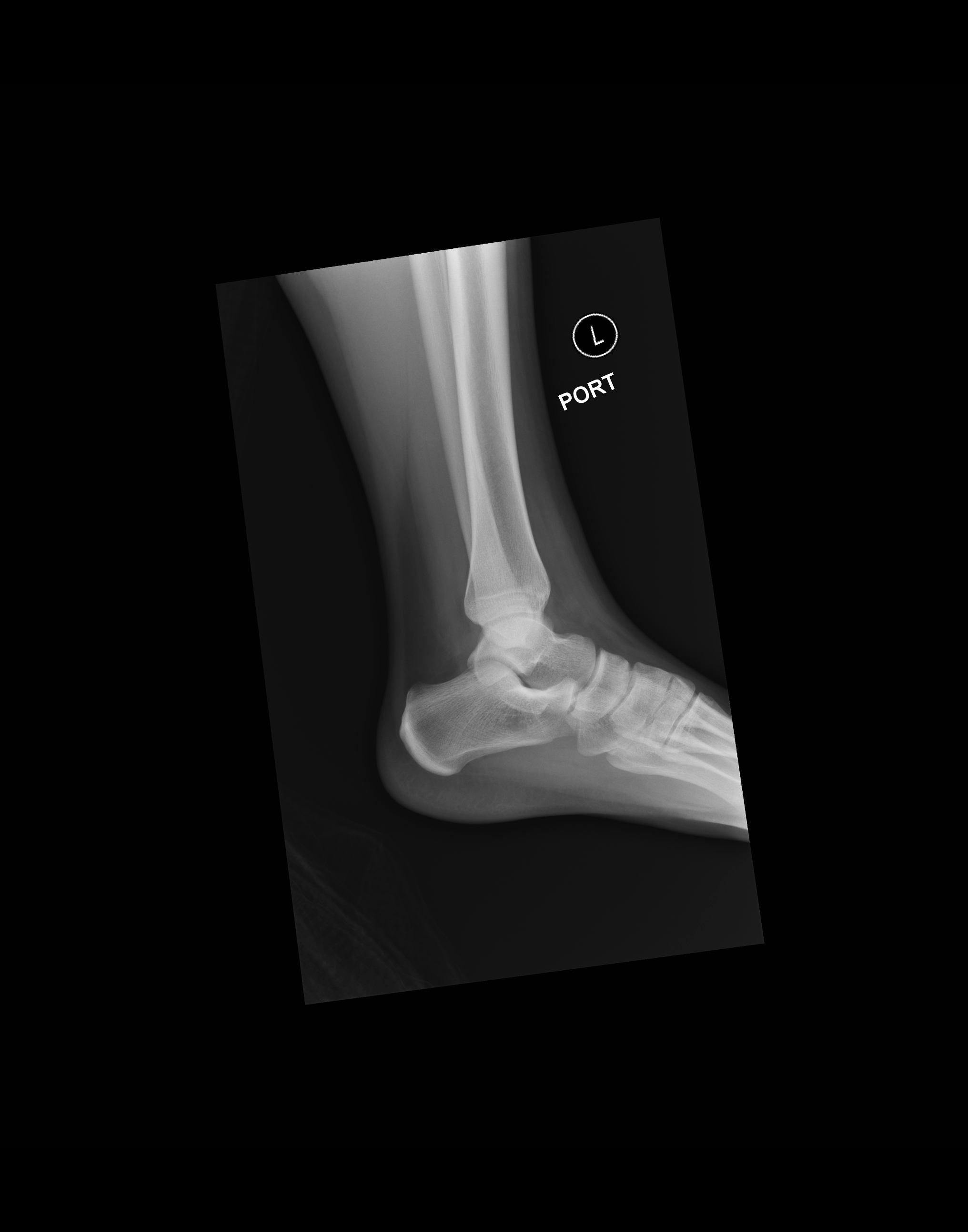 Calcaneal Fracture – TeachMeSurgery – #36
 Radiography techniques of the equine fetlock joint – #37
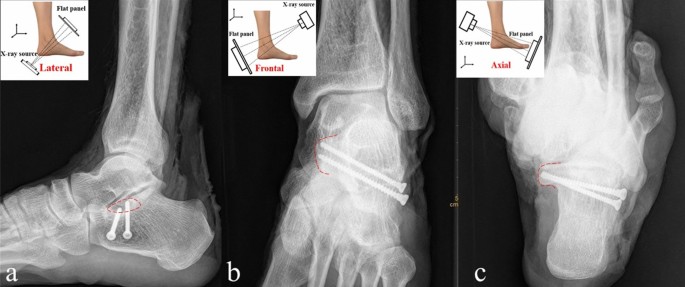 File:Medical X-Ray imaging TKL07 nevit.jpg – Wikimedia Commons – #38
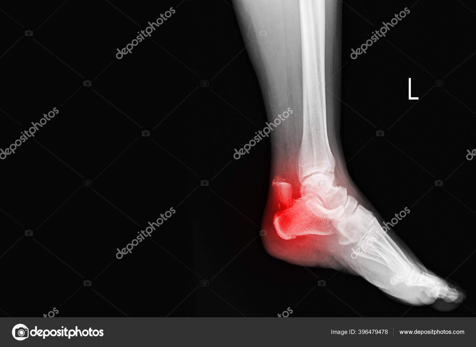 Film ankle X-ray radiograph showing heel bone broken close fracture calcaneus Medical technology and healthcare concept. 27716300 Stock Photo at Vecteezy – #39
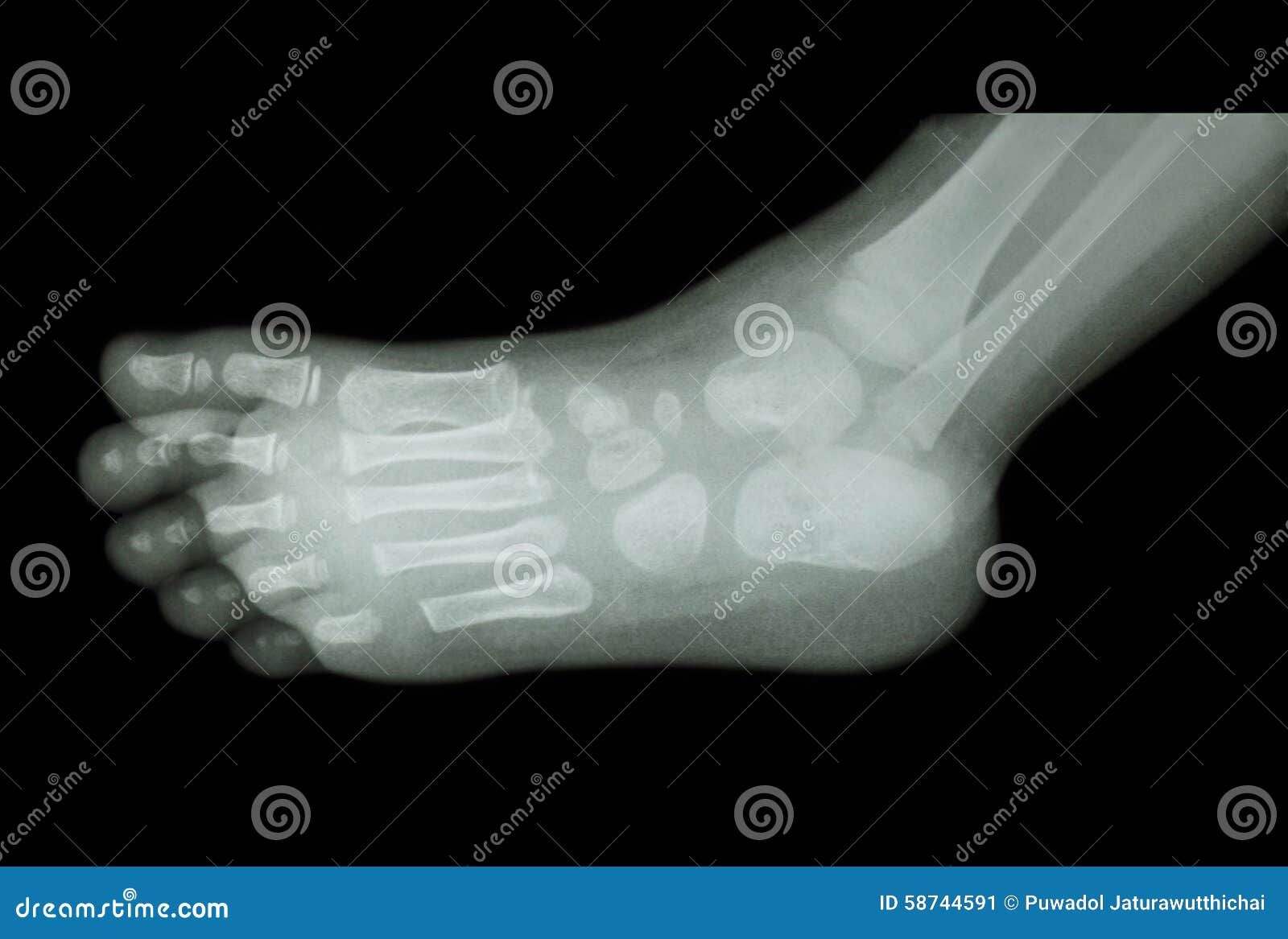 X-ray of the foot oblique view | MyFootShop.com – #40
 Calcaneus as a Rare Location of Solitary Osteochondromas: Two Case Reports in: Journal of the American Podiatric Medical Association Volume 113 Issue 4 (2023) – #41
 e Radiograph (Lateral view of both heels) showing non-union of… | Download Scientific Diagram – #42
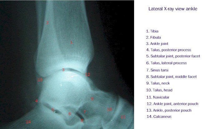 Foot and Heel | Radiology Key – #43
 Peritalar Instability | SpringerLink – #44
 ViewMedica Stock Art: Lateral view of foot – #45
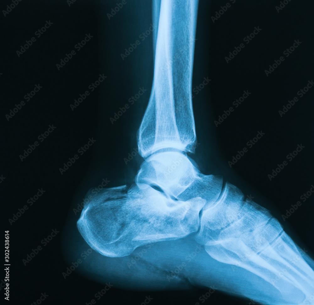
 LATERAL PROJECTIONS : ANKLE XRAY – RadTechOnDuty – #46
 Elbow Joint Section Model at Rs 3770 | Joint Models in Pune | ID: 23461919555 – #47
 Film Ankle X-ray Radiograph Showing Heel Bone Broken 3 Views Close Fracture Calcaneus . Medical Technology and Healthcare Stock Photo – Image of health, orthopedic: 242419886 – #48
 X Ray Ankle Lateral View Stock Photos – Free & Royalty-Free Stock Photos from Dreamstime – #49
 Radiographic Anatomy of the Skeleton: Ankle — Lateral View, Labelled – #50
- x ray heel ap lateral view position
- plantar fasciitis normal heel xray
- calcaneus x ray
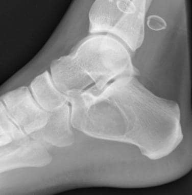
 Calcaneus fracture : r/Radiology – #51
 Off-Track Hooves – Retired Racehorse Project – #52
 Pedobarography shows no differences in gait after talar fractures – IOS Press – #53
- röntgenbild ferse gebrochen
- calcaneus fracture x ray
- normal calcaneus x ray
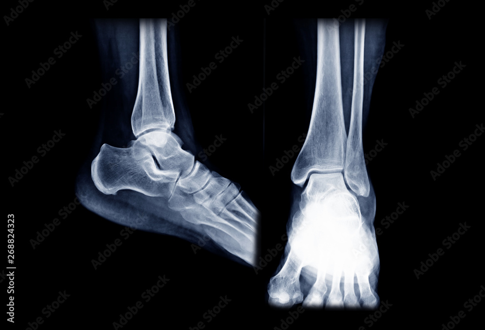 Cureus | Follow the Shoestring: A Unique Case of Bullet Extraction | Article – #54
 Relationship between ankle varus moment during gait and radiographic measurements in patients with medial ankle osteoarthritis | PLOS ONE – #55
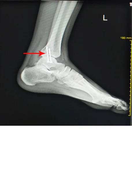 X-ray of the lateral foot | MyFootShop.com – #56
 Premium Photo | Arthritis of ankle. x-ray of foot. lateral view – #57
 File:Radiografia tibiotarsica (caviglia e piede destro) – proiezione laterale.jpg – Wikimedia Commons – #58
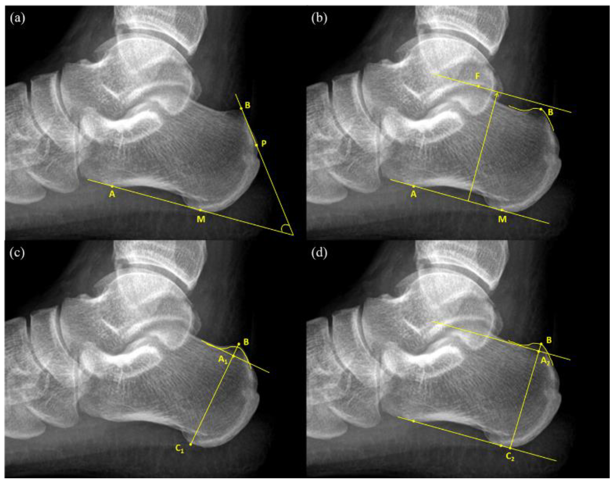 Ray Both Foot Showing Calcaneal Spur Both Side Stock Photo, 48% OFF – #59
 X-ray of the ankle lateral view | MyFootShop.com – #60
- heel ap lat x ray
- heel xray normal
- heel x ray view
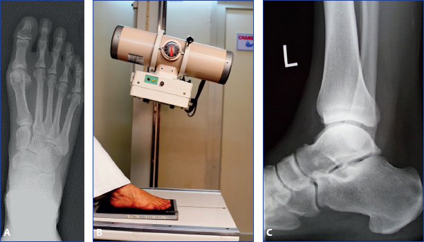 Caudal foot placement superior to toe elevation for navicular palmaroproximal‐palmarodistal‐oblique image quality – Peeters – 2023 – Equine Veterinary Journal – Wiley Online Library – #61
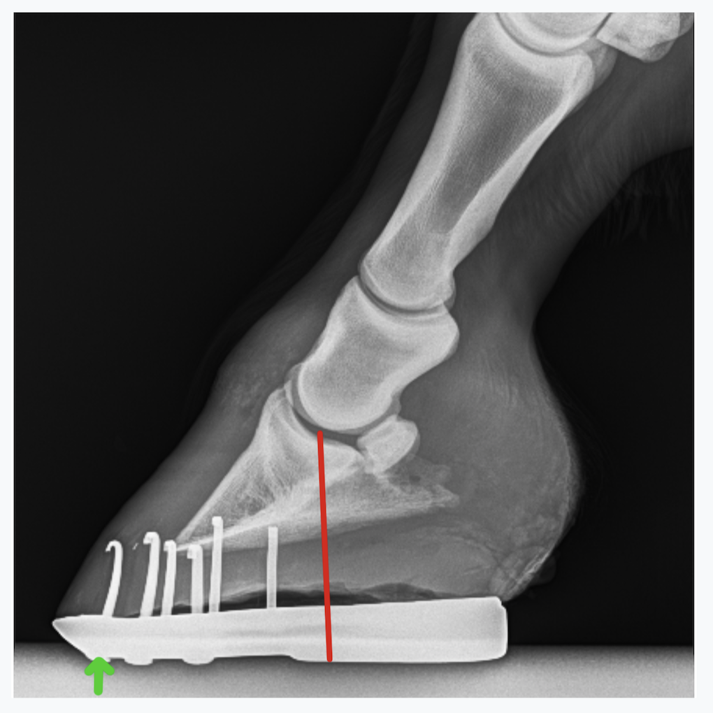 Foot Xray Inverted Lateral View Of Ankle Arthritis Photo Background And Picture For Free Download – Pngtree – #62
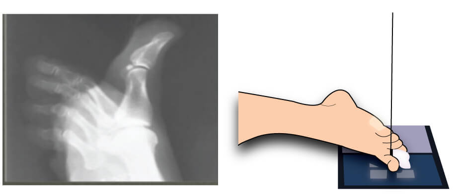 Diagnostic Radiographic Imaging of the Adult Calcaneus – #63
 Shokeen X-ray & Dignostics Centre – #64
 Radiographs of the right foot and ankle. The calcaneus is laterally… | Download Scientific Diagram – #65
 Lateral xray hi-res stock photography and images – Alamy – #66
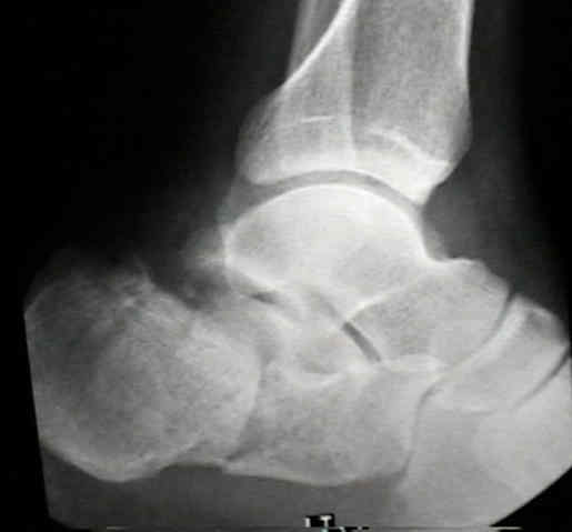 Introduction to Foot and Ankle Radiology Flashcards | Quizlet – #67
- heel spur xray vs normal
- normal foot x ray lateral
- healthy normal heel xray
 Radiography | SpringerLink – #68
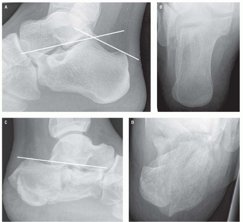 Normal lateral calcaneal radiography | Radiology Case | Radiopaedia.org – #69
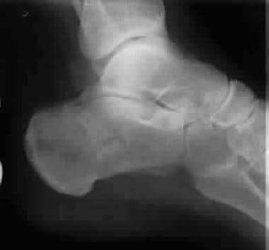 Anatomy of Ankle X-rays – YouTube – #70
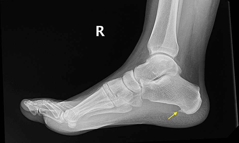 Nail in Foot, X-ray | Stock Image – Science Source Images – #71
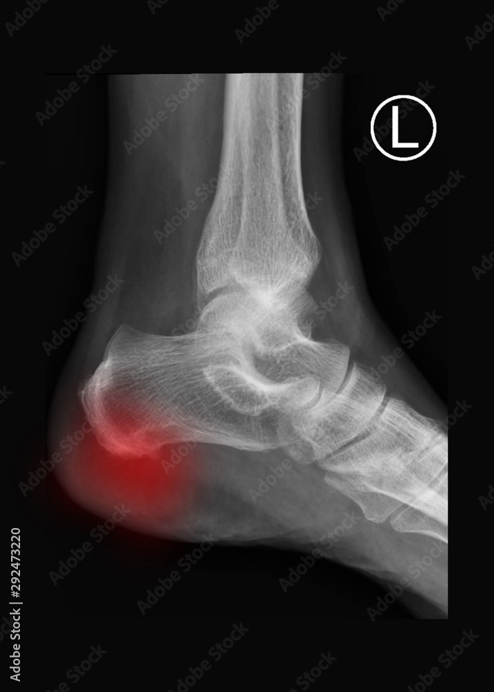 Calcaneal | Orthotics Plus Melbourne – #72
 Tongue-Type Calcaneal Fracture due to a Low-Energy Injury – #73
 OrthoDx: Heel Pain – Clinical Advisor – #74
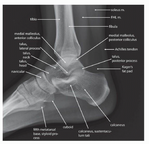 OJHMS | Foot and Ankle Trauma Update 2012 – #75
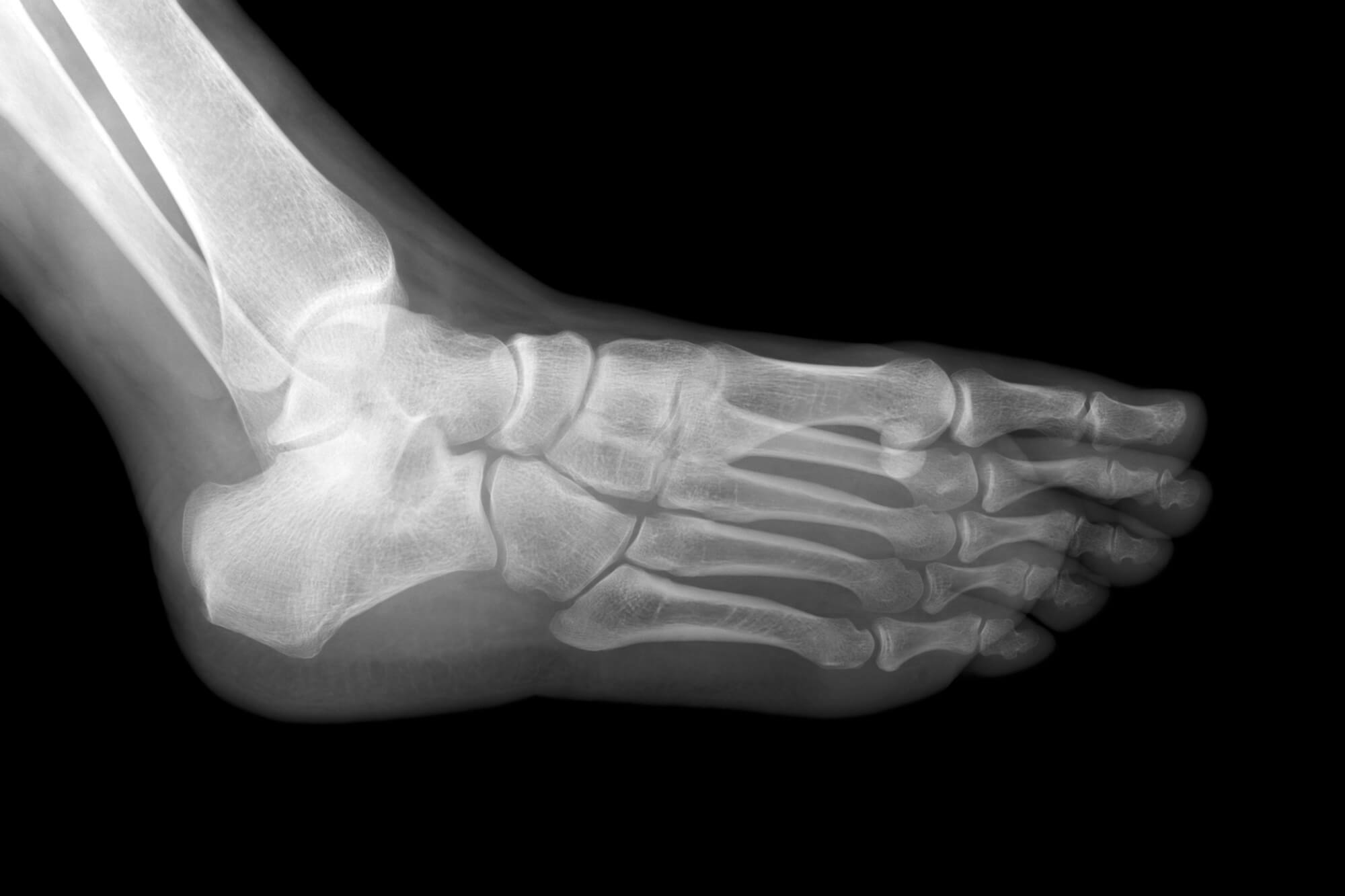 Plantar Fasciitis – Union Spine – #76
 xray of foot by side view Stock Photo | Adobe Stock – #77
 Minimally Invasive Achilles: Haglund’s Syndrome and Endoscopic Calcaneoplasty | OrthoVirginia – #78
 Orthobullets on Instagram: “Can you answer our FREE Question of the Day? A 17-year-old female presents with foot pain associated with footwear. A current lateral radiograph of the affected foot is depicted – #79
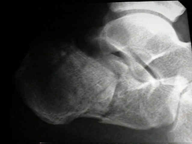 Navicular bone fracture on the x-ray. Trauma from falling of a scooter on the wet road. : r/medizzy – #80
 Ray Right Foot Showing Calcaneal Spur Red Color Copy Space Stock Photo by ©Richmanphoto 242001148 – #81
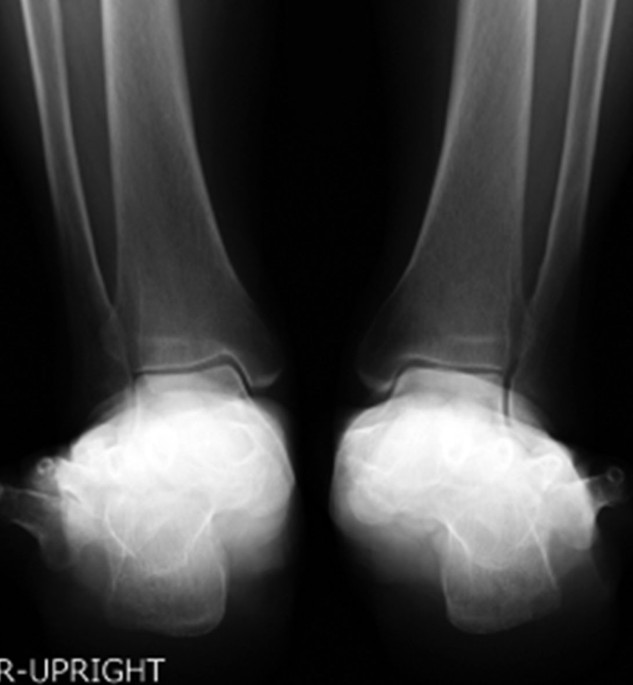 Xray Of The Heel After Calcaneus Fracture Stock Photo – Download Image Now – 2015, Adult, Alloy – iStock – #82
![Figure, Lateral view of the foot. Contributed by Douglas Byerly, MD PhD] - StatPearls - NCBI Bookshelf Figure, Lateral view of the foot. Contributed by Douglas Byerly, MD PhD] - StatPearls - NCBI Bookshelf](https://i.redd.it/i45iegvnxmj71.jpg) Figure, Lateral view of the foot. Contributed by Douglas Byerly, MD PhD] – StatPearls – NCBI Bookshelf – #83
Figure, Lateral view of the foot. Contributed by Douglas Byerly, MD PhD] – StatPearls – NCBI Bookshelf – #83
 Xray Image Of Broken Calcaneus Lateral And Axial View. Stock Photo, Picture and Royalty Free Image. Image 39886937. – #84
 Calcaneal Anastomosis (Left) | Complete Anatomy – #85
 Exostosis – Wikipedia – #86
- both heel lateral x ray
- lateral calcaneus x ray positioning
- foot lateral view positioning
 Case 1) Pre-op X-ray calcaneum lateral view. | Download Scientific Diagram – #87
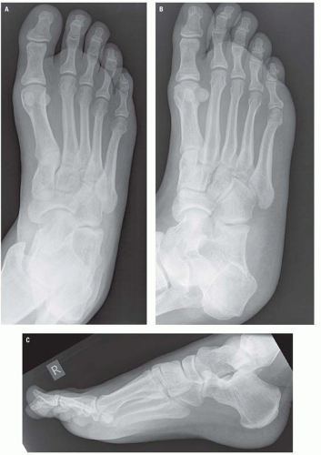 Frontiers | Individual Surgical Treatment of Stage IV Müller-Weiss Disease According to CT/MRI Examination: A Retrospective Study of 12 Cases – #88
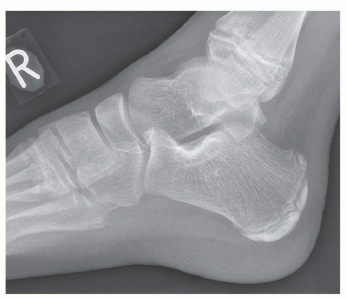 Combining image modalities can enhance diagnosis – Rheuma – #89
 X-ray Image of Ankle, AP and Lateral View. Stock Image – Image of joint, metatarsal: 53839873 – #90
 Case Report of a Tongue-Type Calcaneal Fracture – JETem – #91
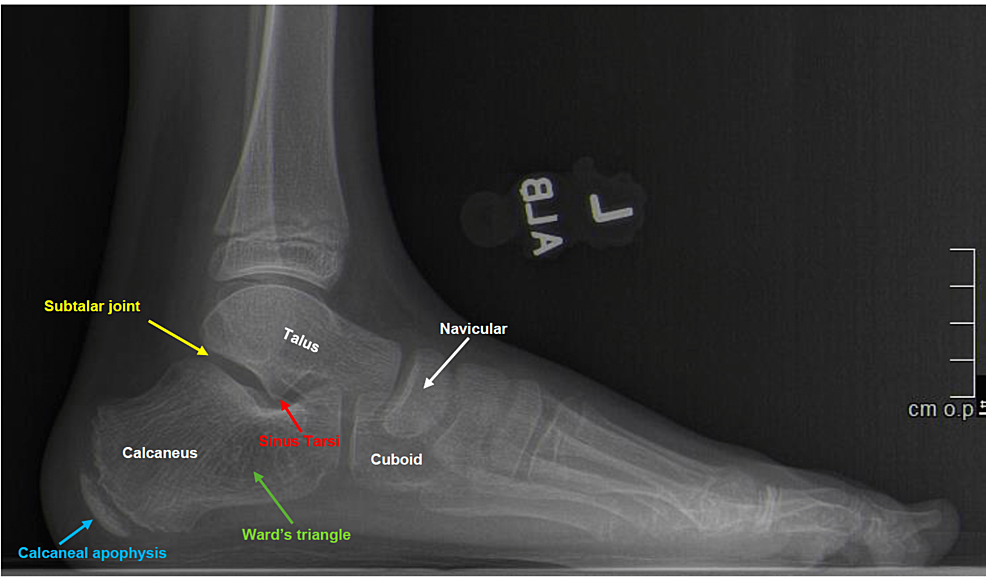 Top Podiatrist Near Me|Family Foot and Leg Center|Best Podiatrist Near Me|Top Doctor Awards|Naples|Estero|Cape Coral|Marco Island|Port Charlotte – #92
- ankle lateral view
- normal foot bones xray
- broken heel xray
 Radiographic imaging of the feet of a 59-year-old man with X-linked… | Download Scientific Diagram – #93
 Calcaneal Spurs, X-ray #2 Galaxy Case by Living Art Enterprises – Science Source Prints – Website – #94
 Pathological ankle fracture due to brown tumour: atypical presentation of low serum vitamin D with normal parathyroid hormone and bone profile | BMJ Case Reports – #95
![Effect of body mass index on soft tissues in adolescents with skeletal class I and normal facial height [PeerJ] Effect of body mass index on soft tissues in adolescents with skeletal class I and normal facial height [PeerJ]](https://pics.craiyon.com/2023-11-23/5x7LtBtXQsiXkfNarHnYAw.webp) Effect of body mass index on soft tissues in adolescents with skeletal class I and normal facial height [PeerJ] – #96
Effect of body mass index on soft tissues in adolescents with skeletal class I and normal facial height [PeerJ] – #96
 Intra-articular Calcaneus Fractures: Current Concepts Review – Paul R. Allegra, Sebastian Rivera, Sohil S. Desai, Amiethab Aiyer, Jonathan Kaplan, Christopher Edward Gross, 2020 – #97
 Xray Image Of Ankle Ap And Lateral View Stock Photo – Download Image Now – Side View, The Human Body, X-ray Image – iStock – #98
 normal lateral x-ray of adult foot Stock Photo – Alamy – #99
 Normal foot x-ray: MedlinePlus Medical Encyclopedia Image – #100
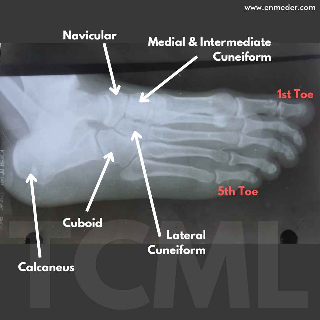
 Tarsal bone – TCML – The Charsi of Medical Literature – #101
 X-ray Right Heel LAT View | Test Price in Delhi | Ganesh Diagnostic – #102
Posts: x ray heel lateral view
Categories: Heels
Author: dienmayquynhon.com.vn
