Update more than 96 heel x ray axial view latest
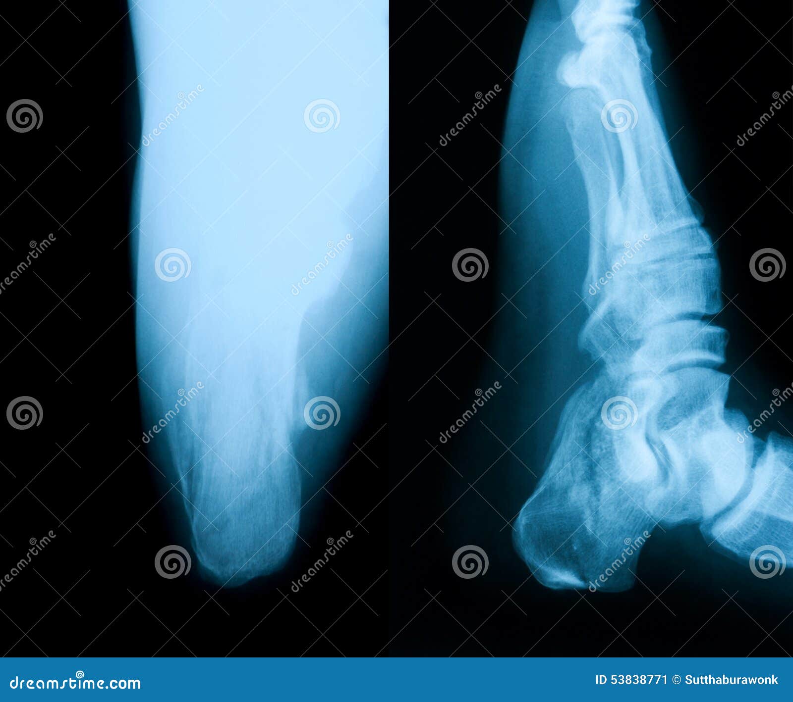
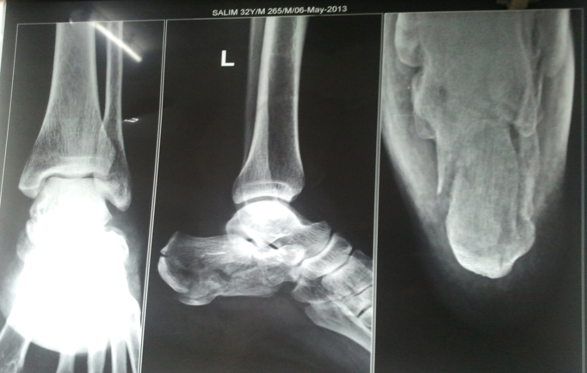
Update images of heel x ray axial view by website dienmayquynhon.com.vn compilation. DIBA: Foot & Ankle Flashcards | Quizlet. The 3D Advantage: Weight Bearing CT vs. X-Ray. X-ray Image of Broken Heel. Stock Image – Image of calcaneus, doctor: 53838771
 Normal Anatomy and Traumatic Injury of the Midtarsal (Chopart) Joint Complex: An Imaging Primer | RadioGraphics – #1
 Diabetic foot syndrome: Charcot arthropathy or osteomyelitis? Part I: Clinical picture and radiography – #2
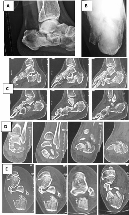 Tarsal coalitions – what you need to know – #3
 Tuberculosis of Foot | Bone and Spine – #4
 Calcaneal spur – Wikipedia – #5
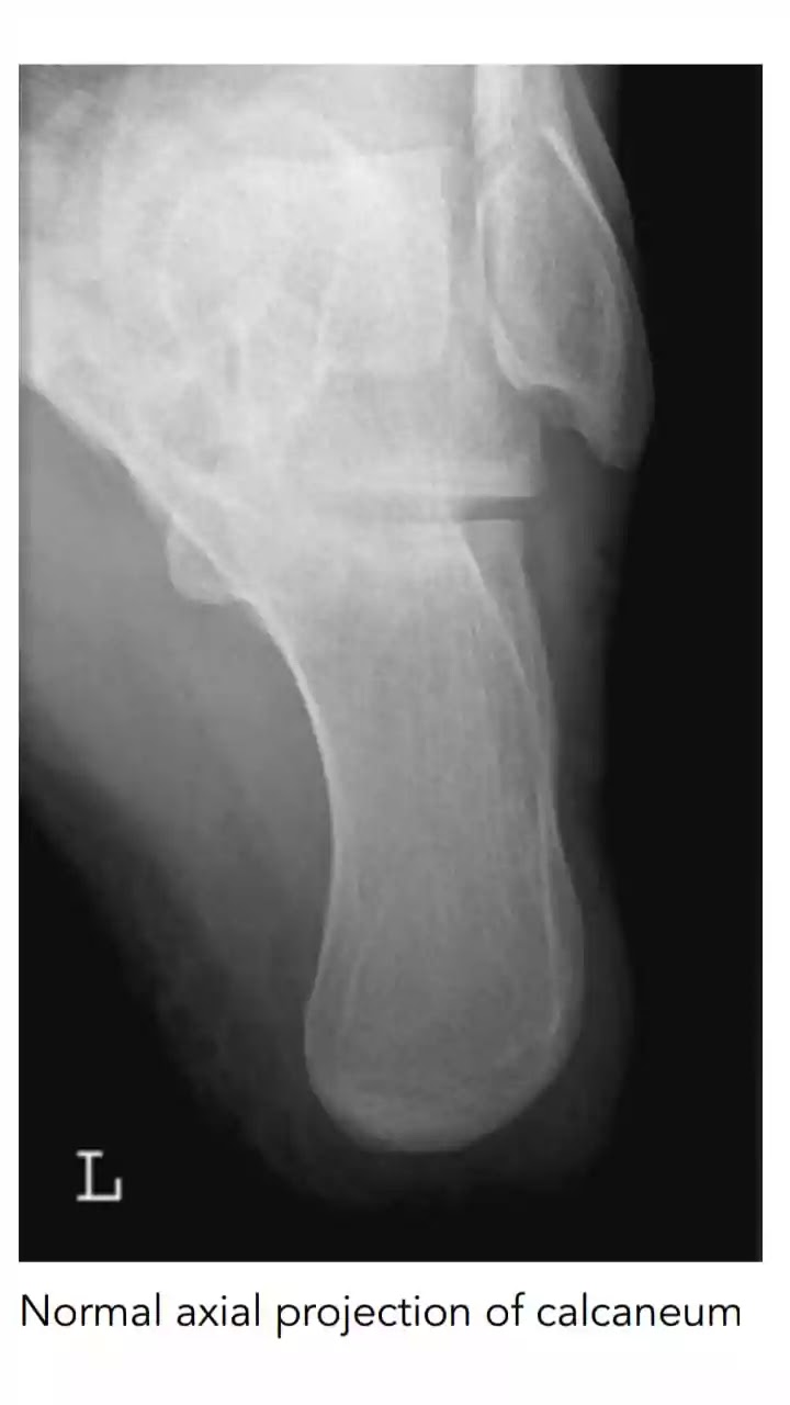
 Normal calcaneum radiographs | Image | Radiopaedia.org – #6
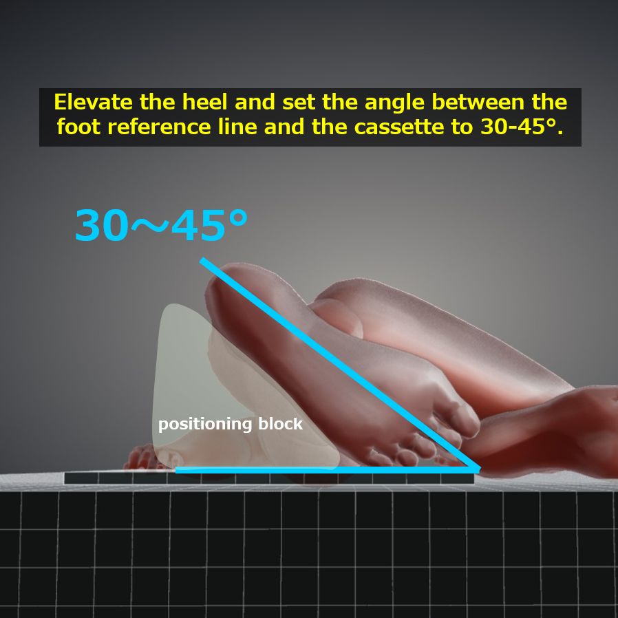 Multiple Reconstructive Osteotomy Treating Malunited Calcaneal Fractures Without Subtalar Joint Fusion – Wang – 2023 – Orthopaedic Surgery – Wiley Online Library – #7
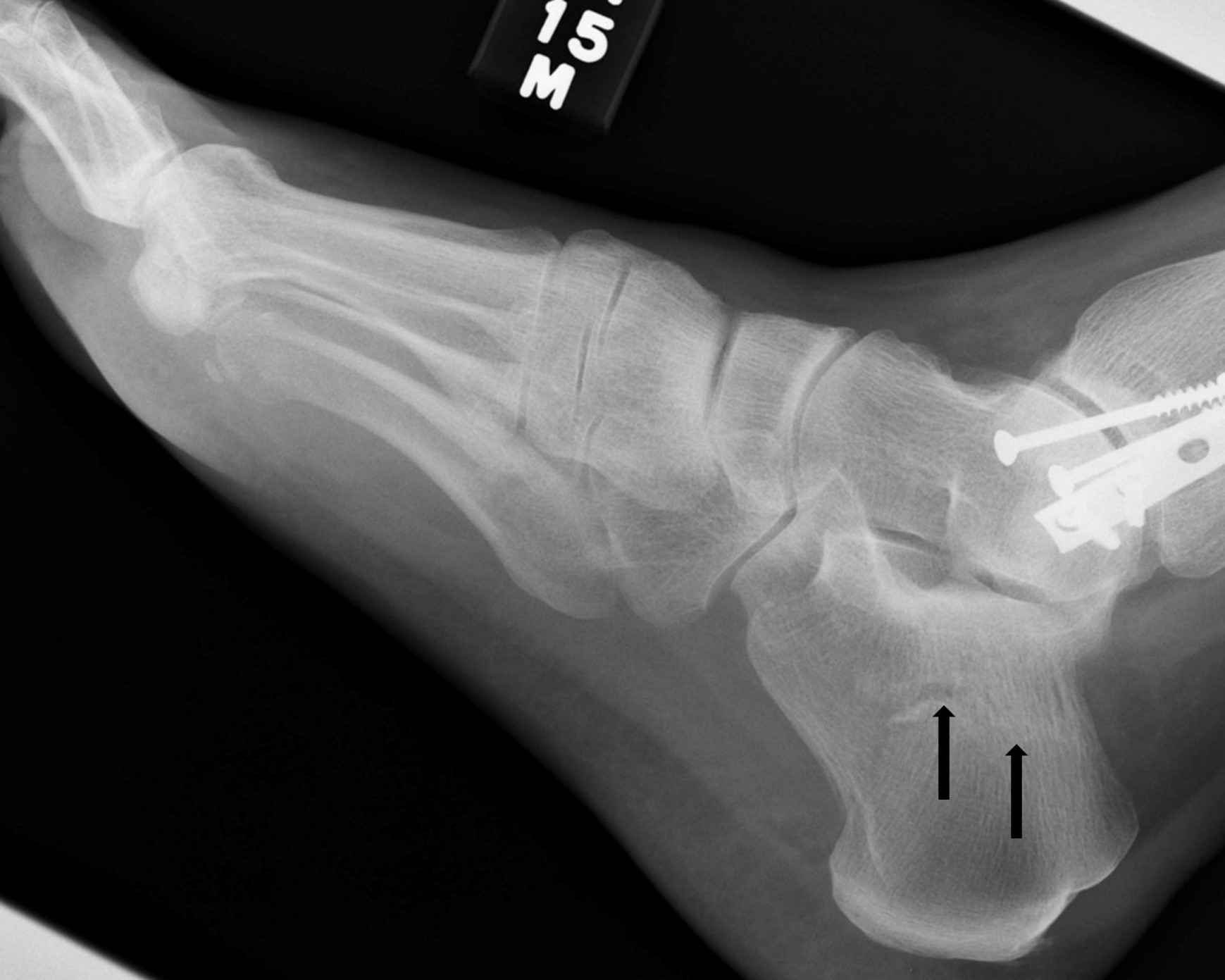 Fractures and dislocations of the tarsal bones | Anesthesia Key – #8
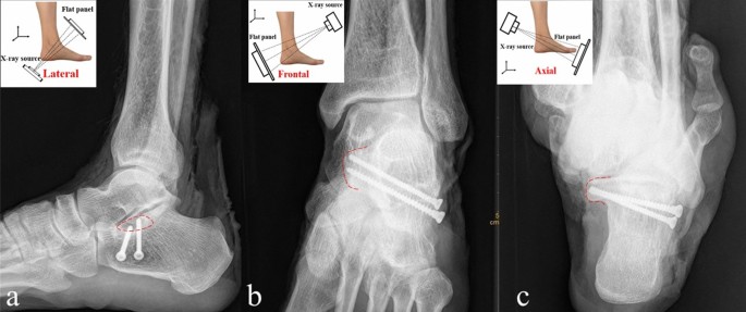 Results and observations in operative treatment of displaced intra- articular calcaneal fractures with use of Calcanail® – #9
 Calcaneus – Reduction & Fixation – ORIF – plate and screw fixation – AO Surgery Reference – #10
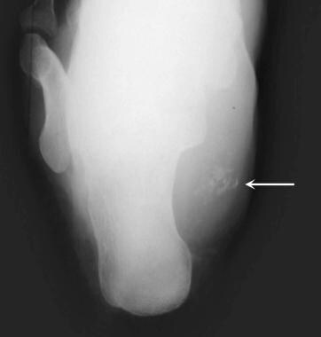
 An Unrecognized Foreign Body Retained in the Calcaneus: A Case Report – #11
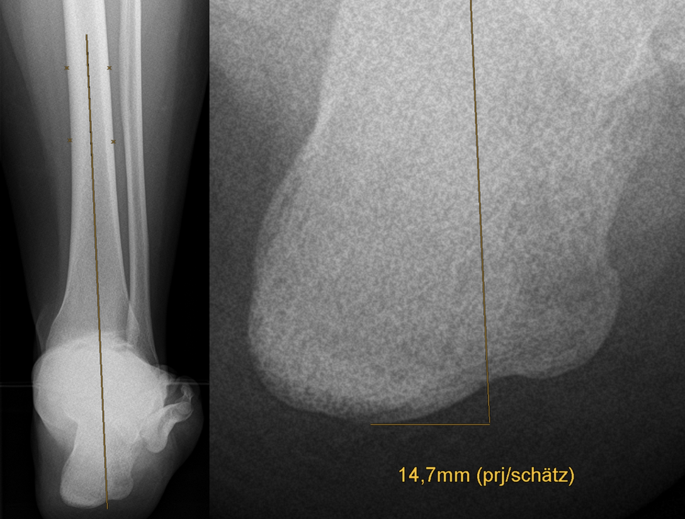
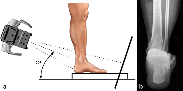
 Measuring standing hindfoot alignment: reliability of different approaches in conventional x-ray and cone-beam CT | Archives of Orthopaedic and Trauma Surgery – #12
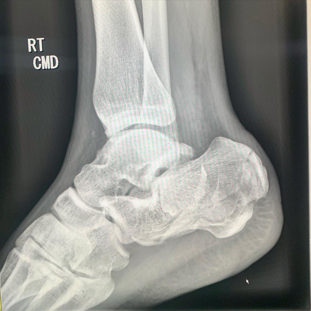 The subtalar joint: A complex mechanism in: EFORT Open Reviews Volume 2 Issue 7 (2017) – #13
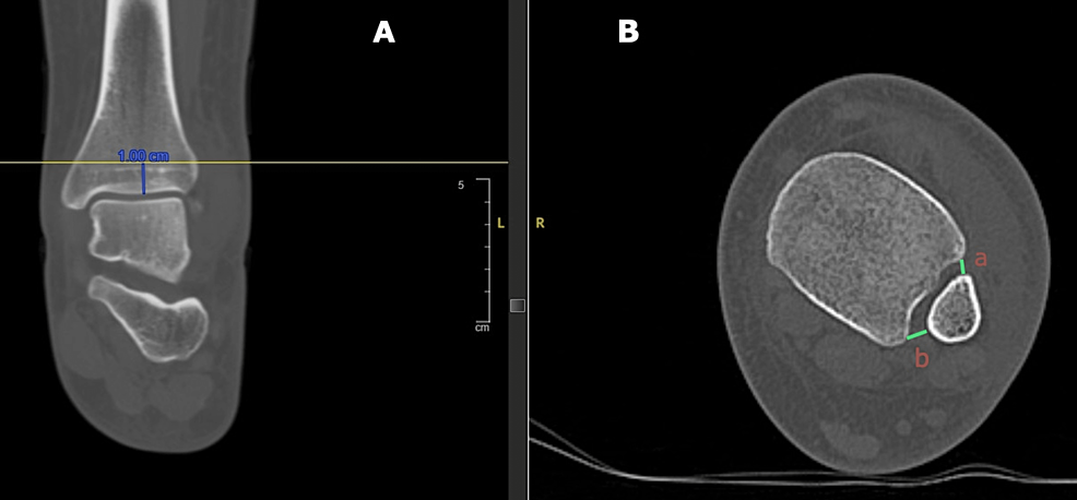 Medical Resources for Charcot Foot & Ankle Specialists – Orthofix – #14
 www.orthojournalhms.org/images/manuscripts/0016_fi… – #15
 Restoring the Anatomy of Calcaneal Fractures: A Simple Technique With Radiographic Review – James M. Cottom, Joseph S. Baker, 2017 – #16
 Calcaneus axial view|Tools for RadTech – #17
- x ray heel ap view positioning
- calcaneus ap position
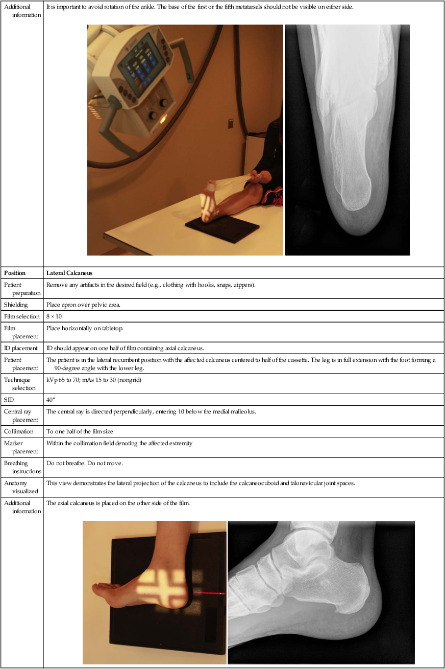
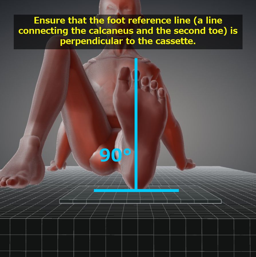
- calcaneal axial view positioning
 Case Study: Injectable Bone Cement in Treatment of ORIF Calcaneal Fractures – #18
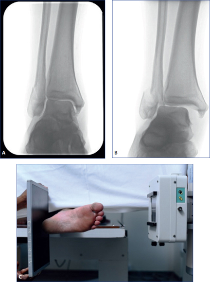 Acute Fractures and Dislocations of the Ankle and Foot in Children – #19
 A) Lateral X-ray of calcaneus, obvious displacement of calcaneal… | Download Scientific Diagram – #20
 Radiographic Assessment of Pediatric Foot Alignment: Review | AJR – #21
 X-ray Right Calcaneum AP-Axial | Test Price in Delhi | Ganesh Diagnostic – #22
 Congenital Variations of the Peroneal Tendons | SpringerLink – #23
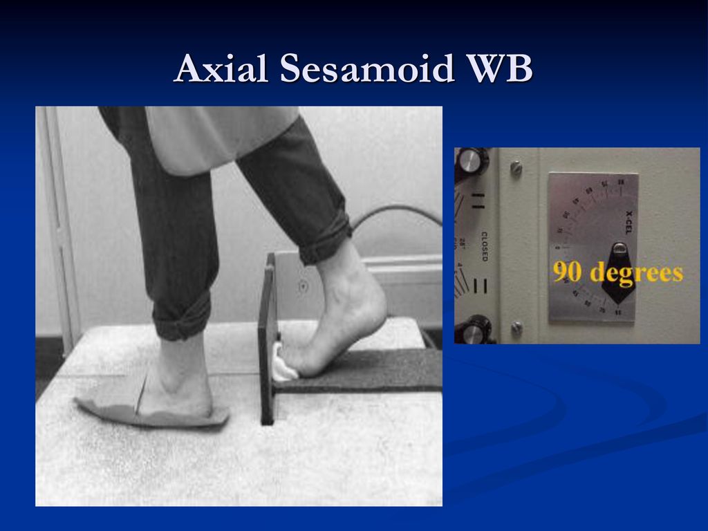
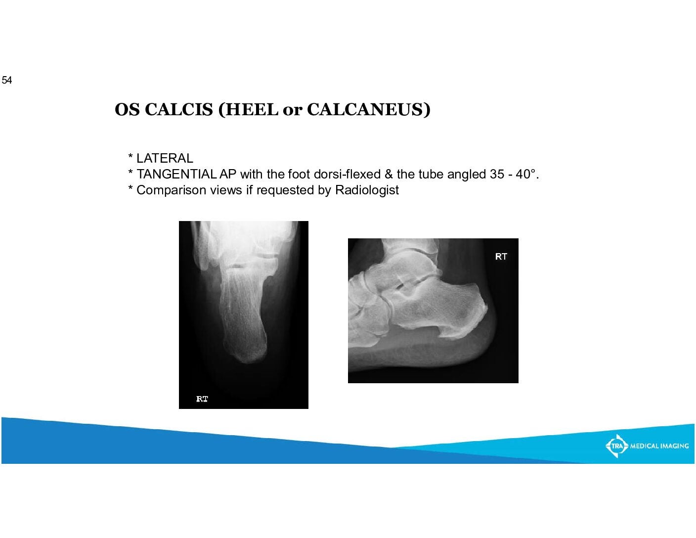
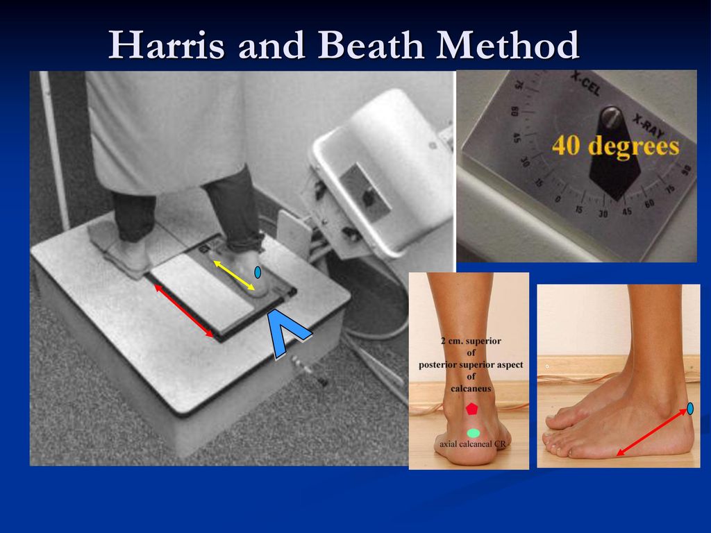
 Radiographic Positioning of the Foot & Ankle – ppt download – #24
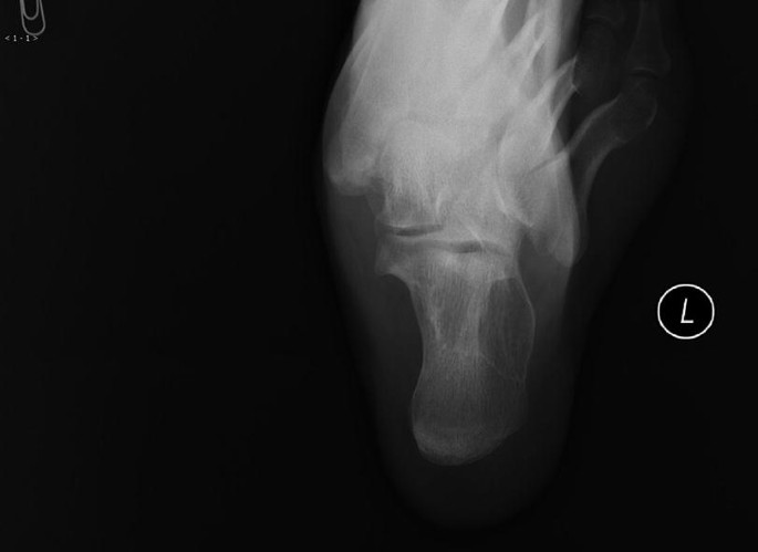 Ankle & hindfoot | Radiology Key – #25
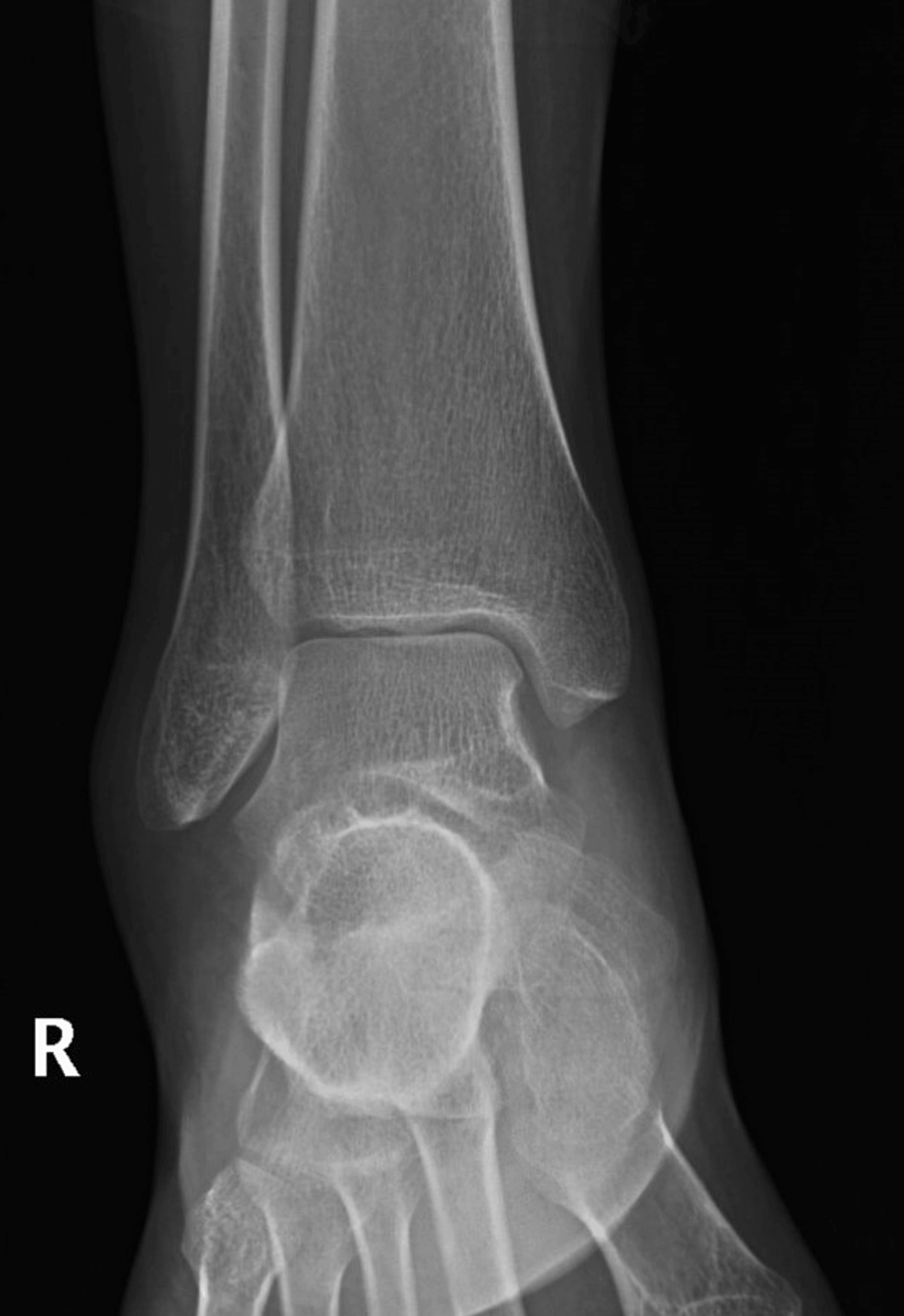 A Slowly Healing Leg Wound – Journal of Urgent Care Medicine – #26
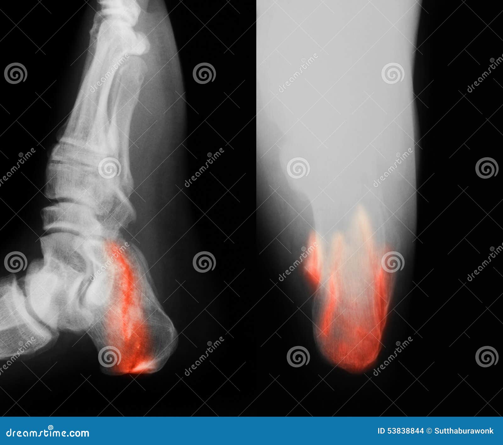 X-ray Left Calcaneum AP Axial | Test Price in Delhi | Ganesh Diagnostic – #27
 Periarticular opening wedge osteotomy for severe valgus deformity and associated rearfoot tarsal coalitions – #28
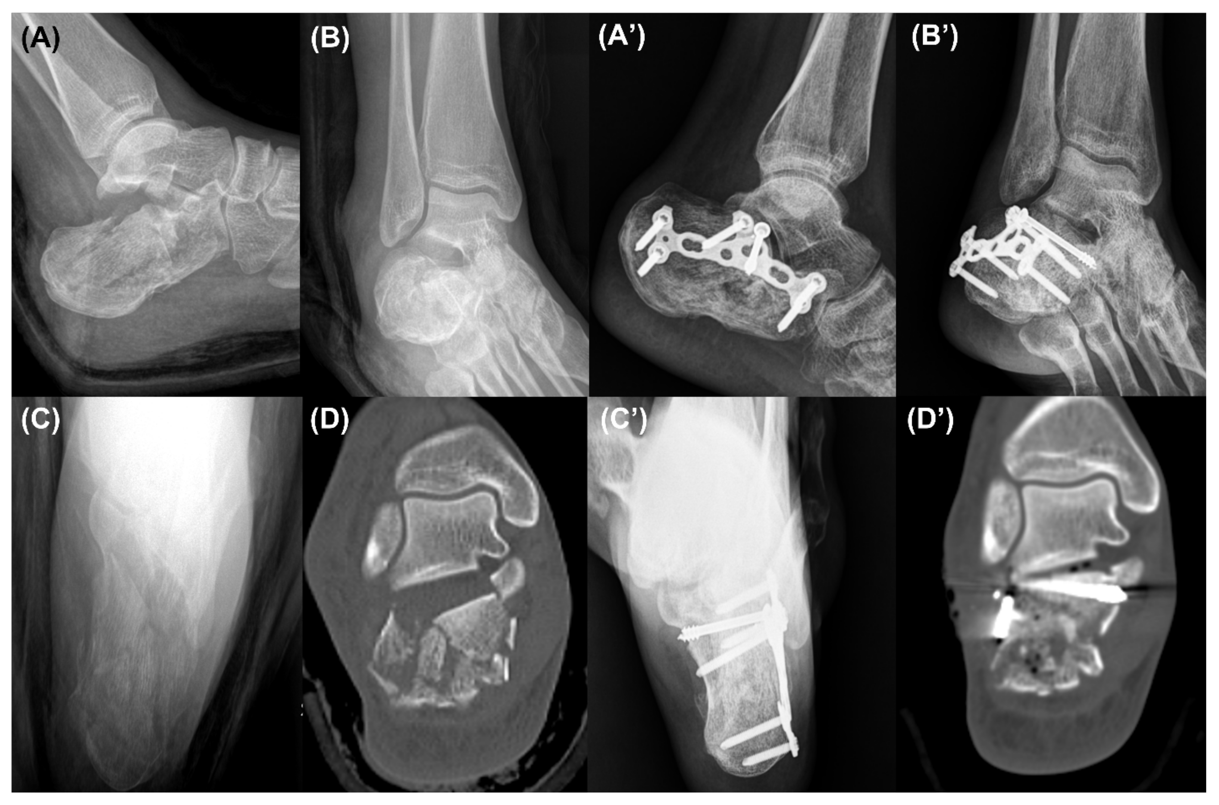
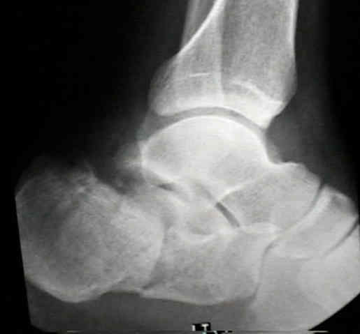 Postoperative radiographs (A, lateral view; B, axial view) and CT… | Download Scientific Diagram – #29
 Montefiore Rehabilitation Medicine on X: “Calcaneus fracture make up 50-60% of tarsal fractures, usually resulting from a high impact axial trauma (AKA falling from a ladder or MVA) Initial clinical presentation will – #30
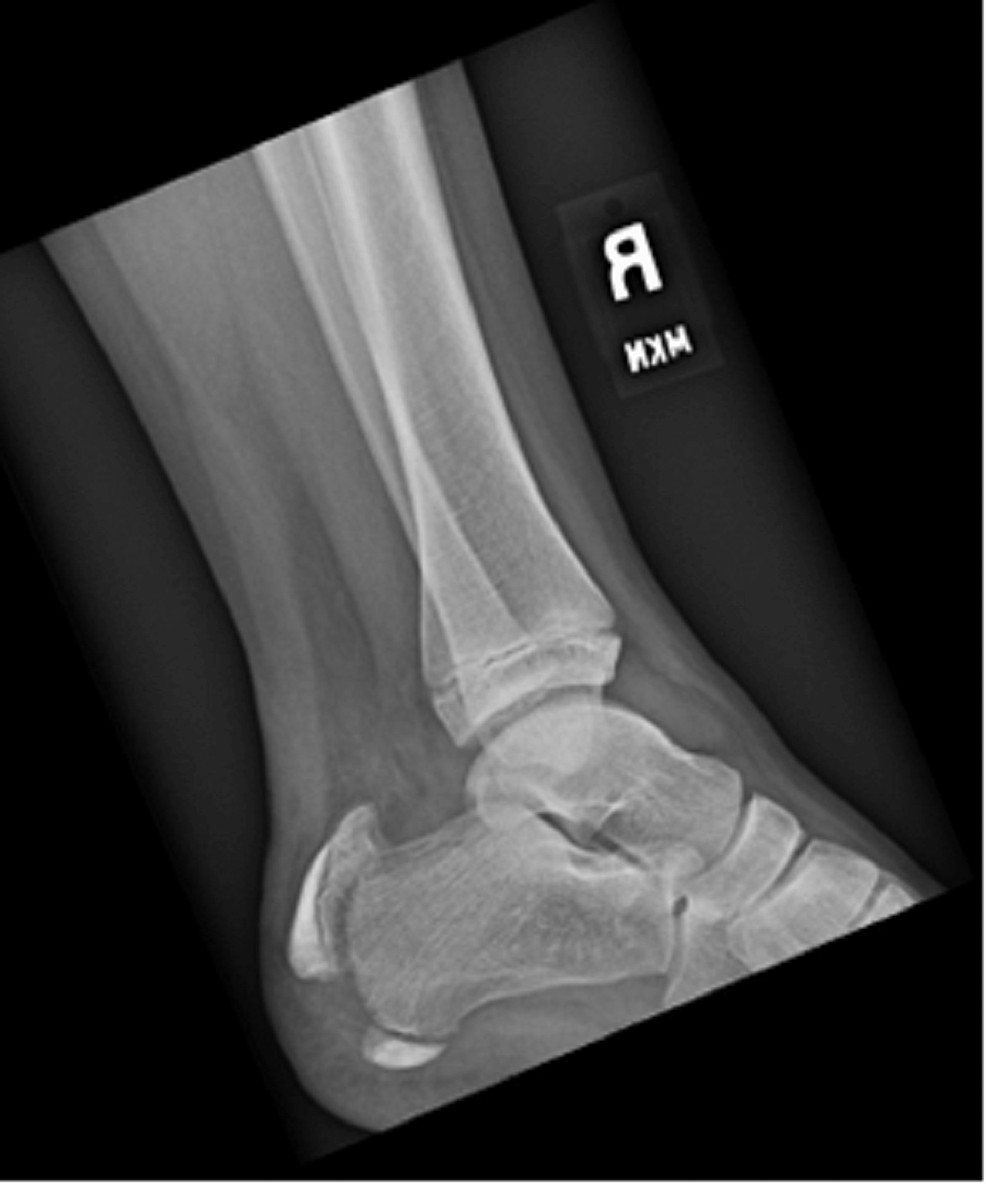 Treatment of the Accessory Navicular – #31
 How To Evaluate And Treat Calcaneal Fractures – #32
 Figure 7 from Angle and Base of Gait Long Leg Axial and Intraoperative Simulated Weightbearing Long Leg Axial Imaging to Capture True Frontal Plane Tibia to Calcaneus Alignment in Valgus and Varus – #33
- harris view ankle
- calcaneus axial view anatomy
- x ray calcaneus lateral view
- calcaneus axial position
- sustentaculum tali xray
 Knee Hyperextension in Stance Phase of Gait in Hemiparesis Factors and Their Management: An Exploratory Case Series – #34
 Hindfoot Alignment Measurements: Rotation-Stability of Measurement Techniques on Hindfoot Alignment View and Long Axial View Rad – #35
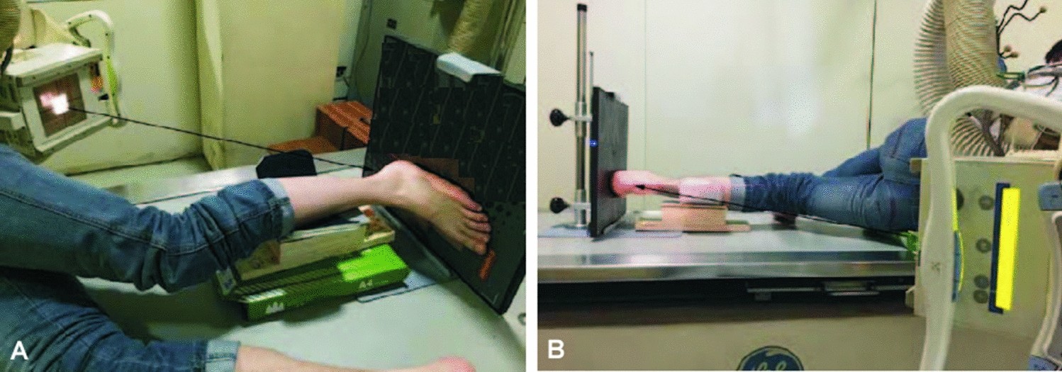 Basics – X-Rays Flashcards | Quizlet – #36
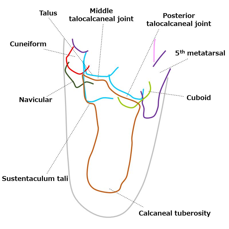 Intra-articular Calcaneal Fractures | SpringerLink – #37
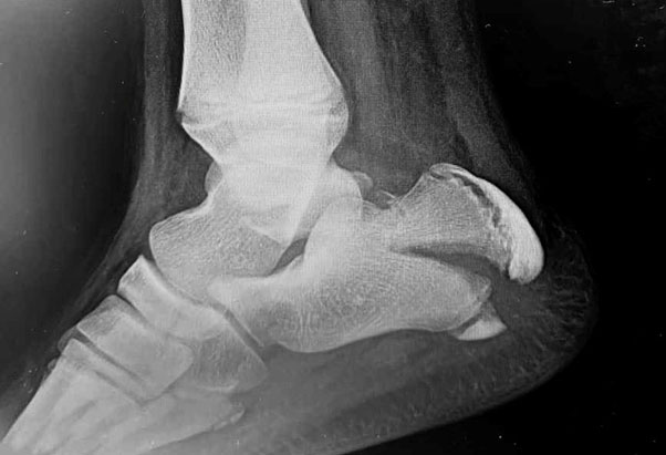 UCSD Musculoskeletal Radiology – #38
 SURGICAL PLANNING IN MIDDLE FACET TALOCALCANEAL COALITION – #39
 Calcaneal Intraosseous Lipoma treated with External Fixation: A case report and review of the literature | The Foot and Ankle Online Journal – #40
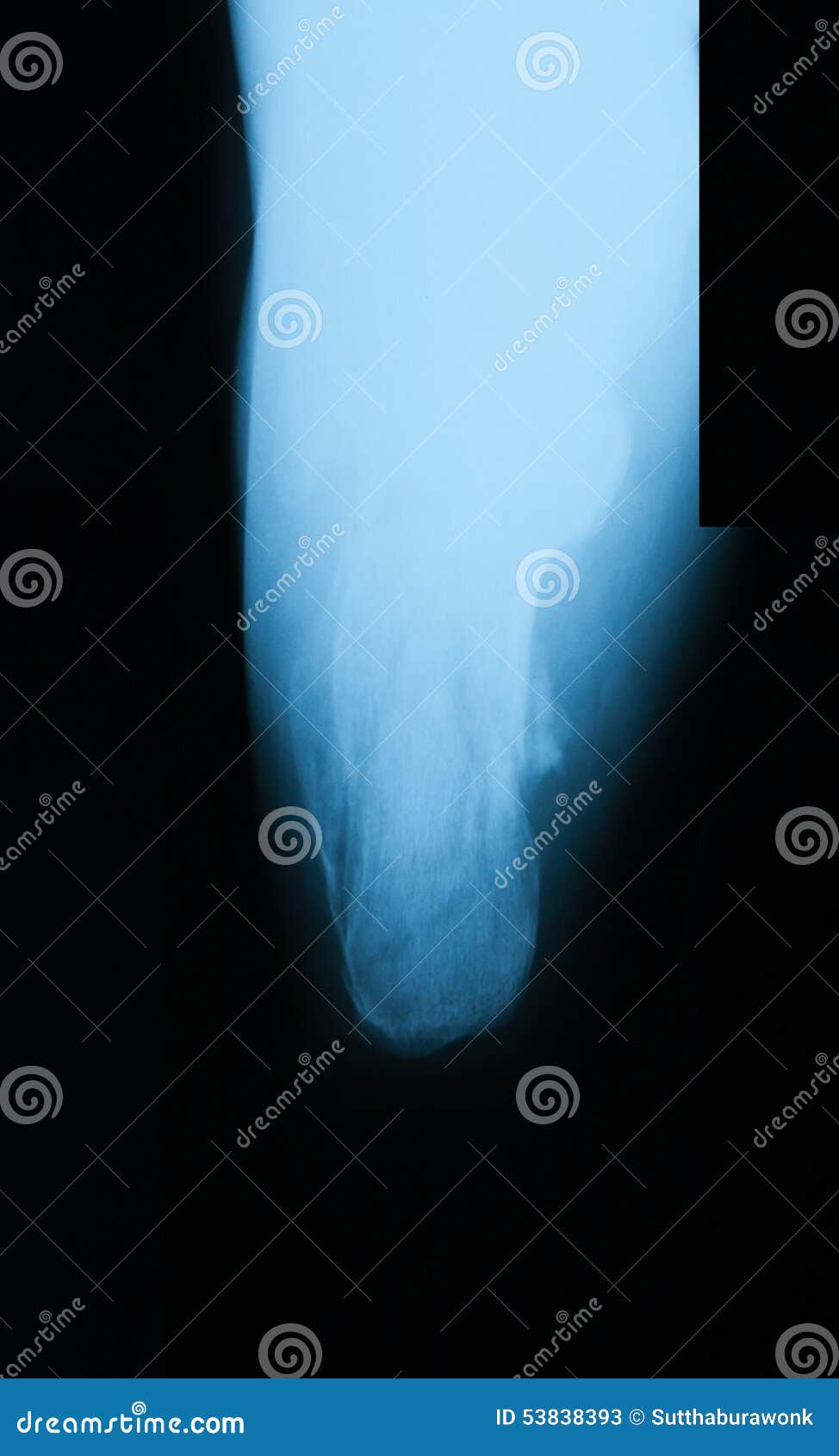
 X-ray image of calcaneus. stock image. Image of foot – 53838393 – #41
 Mri heel hi-res stock photography and images – Alamy – #42
 22 – It’s Not Always Calcaneal Apophysitis – #43
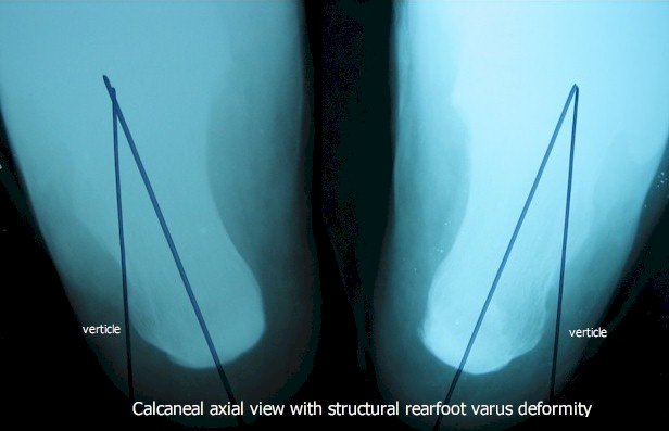
 Measuring hindfoot alignment radiographically: the long axial view is more reliable than the hindfoot alignment view | Skeletal Radiology – #44
 Minimally Invasive Reduction of Intraarticular Calcaneal Fractures With Percutaneous Fixation Using Cannulated Screws Versus Kirschner Wires: A Retrospective Comparative Study – Ahmed Shams, Osama Gamal, Mohamed Kamal Mesregah, 2023 – #45
 Fractures and dislocations of the tarsal bones (Chapter 13) – Broken Bones – #46
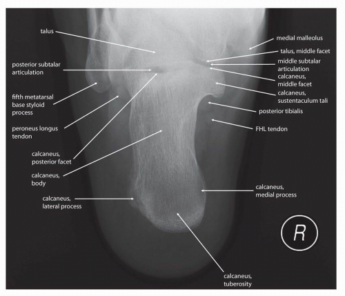 Figure 1 from The Calcaneus Fracture of Joint Depression Type with Lateral Subtalar Dislocation (A Case Report) | Semantic Scholar – #47
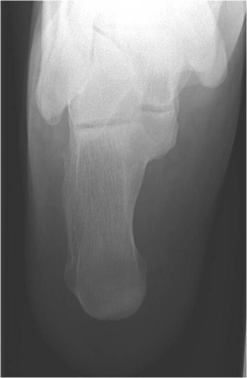
 Metastatic Disease: Case 11 | SpringerLink – #48
 John Sessions, DPM, PhD, Travis McKeller, DPM, Alden L. Simmons, DPM, Douglas Murdoch, DPM, Christopher Browning, DPM, FACFAS – #49
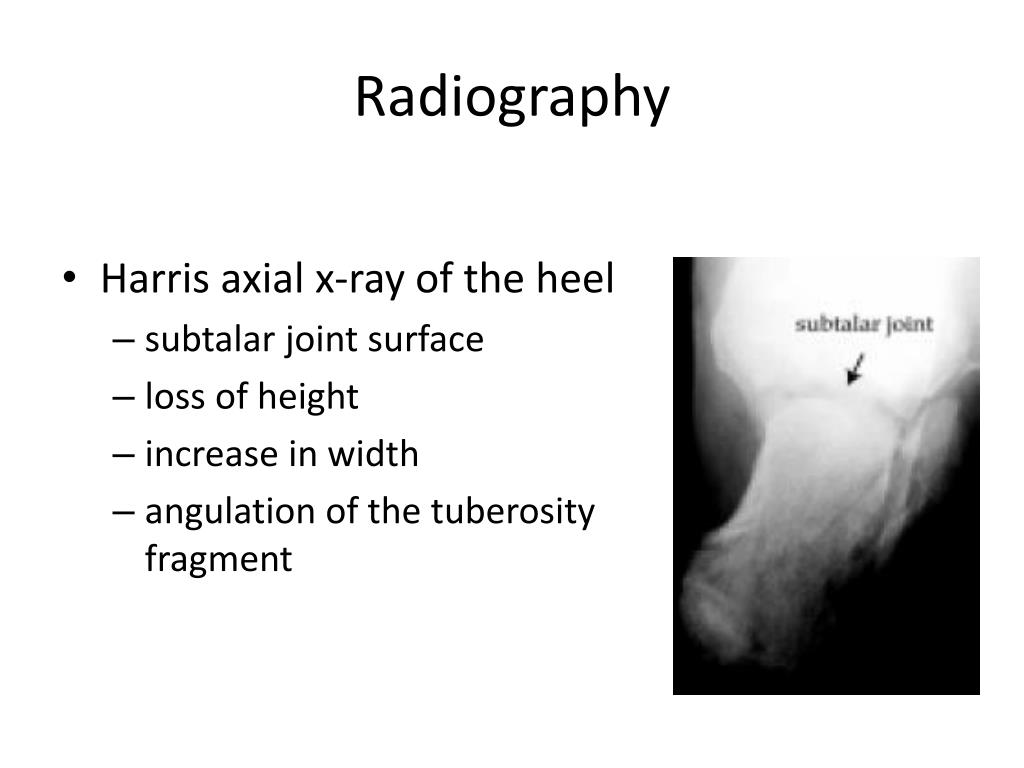
![Figure, Right foot Radiograph Harris view...] - StatPearls - NCBI Bookshelf Figure, Right foot Radiograph Harris view...] - StatPearls - NCBI Bookshelf](https://samarpanphysioclinic.com/wp-content/uploads/2023/05/Calcaneal-fracture-1200x675.webp) Figure, Right foot Radiograph Harris view…] – StatPearls – NCBI Bookshelf – #50
Figure, Right foot Radiograph Harris view…] – StatPearls – NCBI Bookshelf – #50
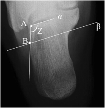 Degenerative Cervical Myelopathy: Natural History, clinical presentation, current diagnosis and treatment update. | Maeda | International Journal of Orthopaedics – #51
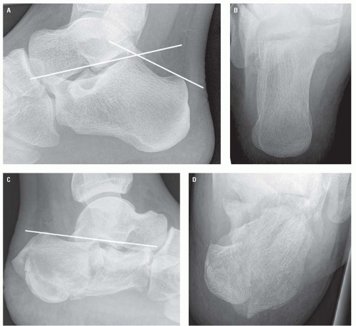 Inaccuracy of Forefoot Axial Radiographs in Determining the Coronal Plane Angle of Sesamoid Rotation in Adult Hallux Valgus Deformity: A Study Using Weightbearing Computed Tomography in: Journal of the American Podiatric Medical – #52
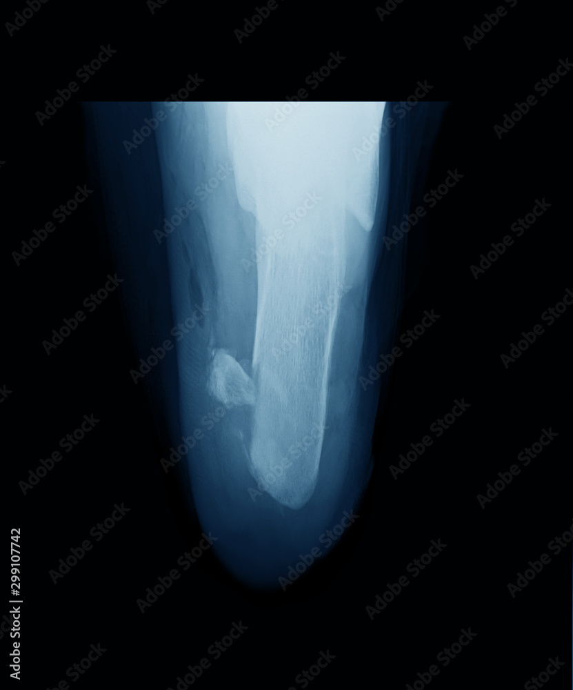 Figure 2 from The Calcaneus Fracture of Joint Depression Type with Lateral Subtalar Dislocation (A Case Report) | Semantic Scholar – #53
 Amazon.com: Colortrieve X-Ray Positioner – Podiatry Axial/Sesamoid Weight Bearing – 13″ x 8″ x 4″ (Closed Cell) : Industrial & Scientific – #54
- axial calcaneus
- calcaneus axial x ray position
- calcaneus fracture axial view
 Normal calcaneal radiographs – paediatrics | Image | Radiopaedia.org – #55
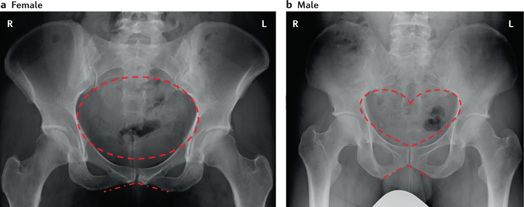
 X-Knee – #56
 Ortho Blog – CMC COMPENDIUM – #57
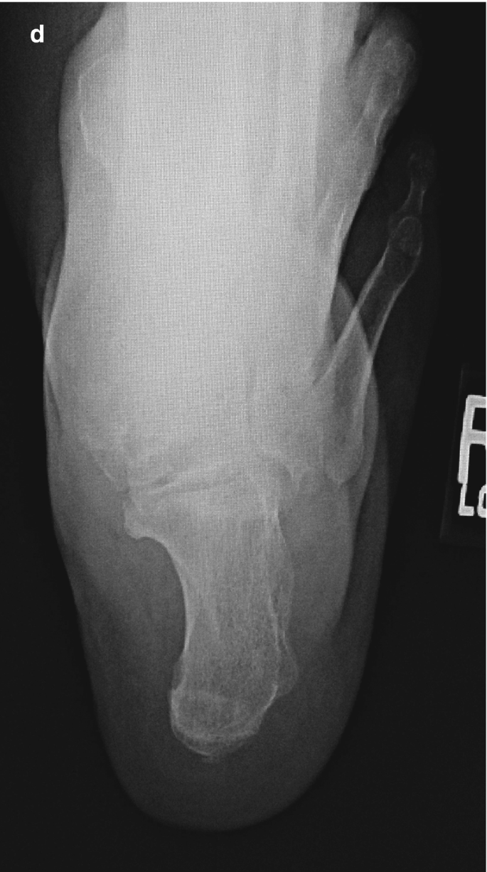 Axial hip view | X ray, Hips, Axial – #58
 Operative Versus Non-Operative Treatment for Closed Displaced intra-articular Fractures of the Calcaneum Introduction – #59
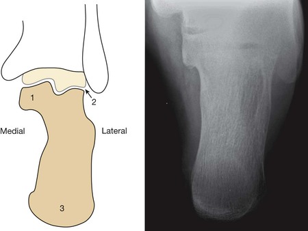 Does axial view still play an important role in dealing with calcaneal fractures? | BMC Surgery | Full Text – #60
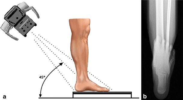 X-ray image of axial calcaneus (heel) showing achilles tendon • wall stickers injury, fracture, disease | myloview.com – #61
- x ray heel ap lateral view position
- saltzman view
- harris axial view position
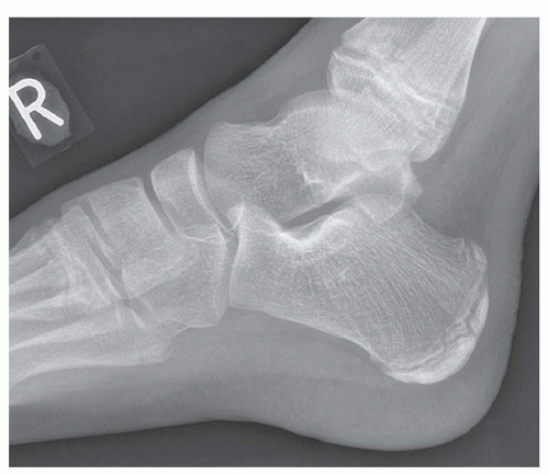 Elbow joint # Axial view X-Ray # Patient position for X-Ray # By BL Kumawat # – YouTube – #62
 Xray Image Broken Calcaneusheel Axial View Stock Photo 269956955 | Shutterstock – #63
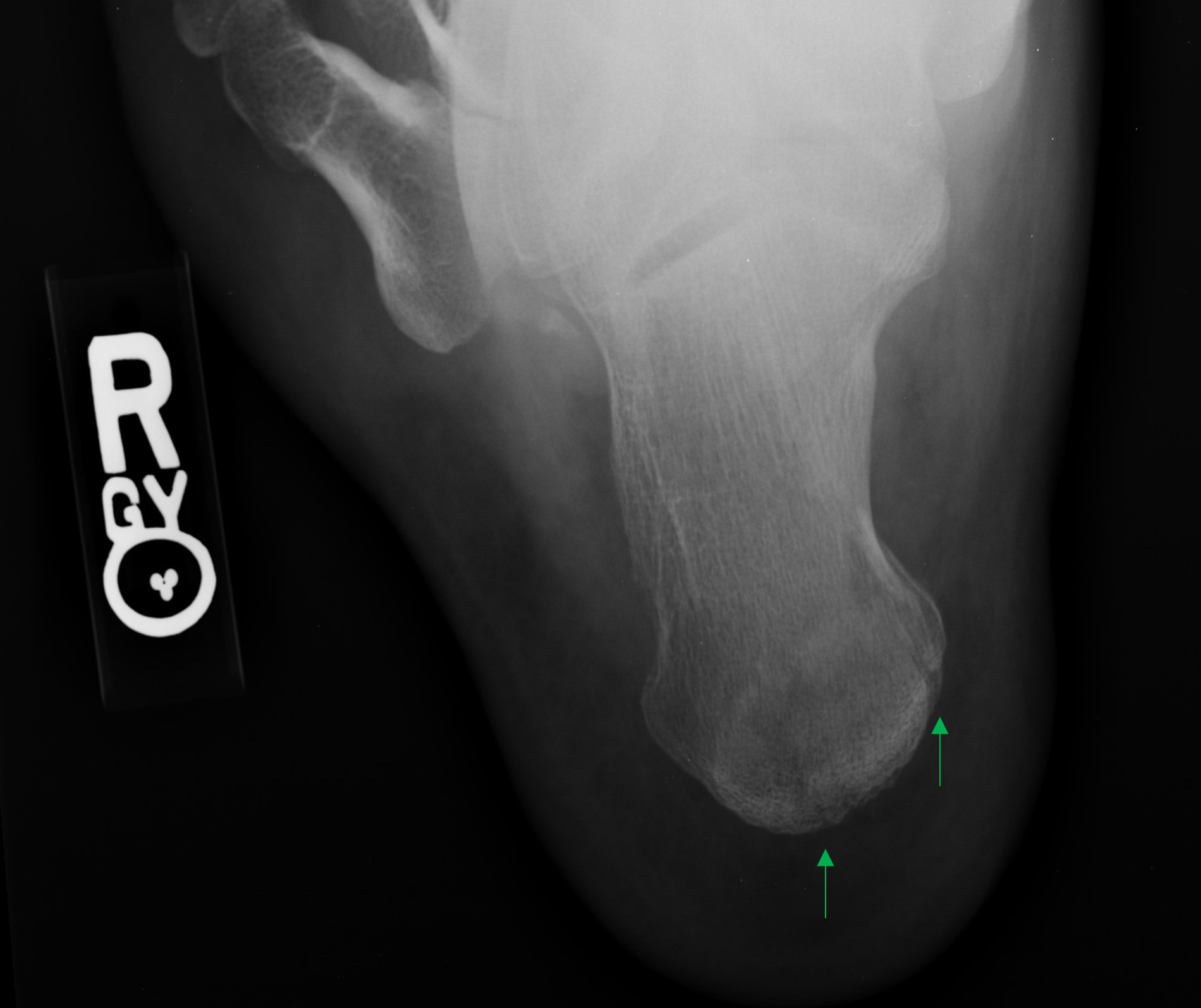
 200+ X Ray Of Human Broken Heel Foot Bone Stock Photos, Pictures & Royalty-Free Images – iStock – #64
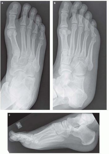
 Foot Case 4 Additional Images: Orthopedic Teaching: Feinberg School of Medicine – #65
 MRI of Heel Pain | AJR – #66
 Case Report A Novel Minimally Invasive Reduction Technique by Balloon and Distractor for Intra-Articular Calcaneal Fractures: A – #67
 Inter- and Intra-observer Reliability of a New Classification System for Calcaneus Fracture Malunions: The ADEINS Classification | Indian Journal of Orthopaedics – #68
 Full article: MRI of the Achilles tendon: A comprehensive review of the anatomy, biomechanics, and imaging of overuse tendinopathies – #69
 Review of Calcaneal Osteotomies Fixed With a Calcaneal Slide Plate – Erin K. Haggerty, Stephanie Chen, David B. Thordarson, 2020 – #70
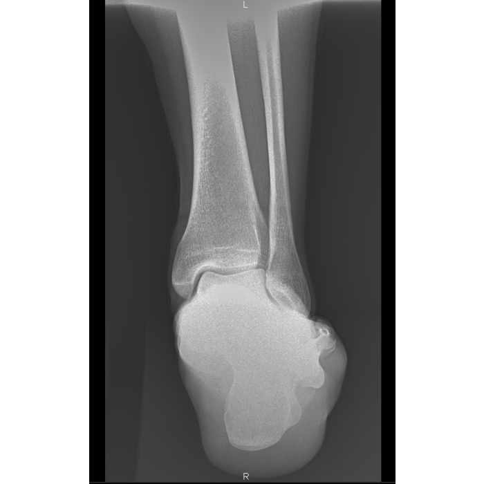 Value of modified axial review radiograph in diagnosing calcaneal fractures | Scientific Reports – #71
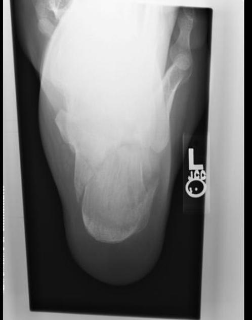 From the EMDaily Archives: What’s the Diagnosis? By Dr. Rebecca Fieles – – #72
 Orthopedic Pitfalls in the ED: Calcaneal Fractures – #73
- harris beath view vs calcaneal axial
- axial calcaneal xray
- harris view
- calcaneal view position
- lateral calcaneus positioning
- calcaneus lateral view position
 Buy Podiatry Axial & Sesamoid Sponge for only $113 at Z&Z Medical – #74
 Tongue-Type Calcaneal Fracture due to a Low-Energy Injury – #75
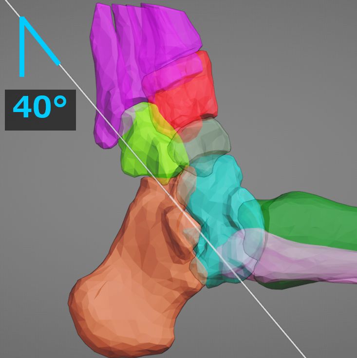 Radiographic evaluation: Harris heel view. Left is normal with parallel… | Download Scientific Diagram – #76
 Is Heel Pain keeping You from Being Productive at Work and Fully Enjoying Life? – Impact Health Niagara – #77
 Posterior Subtalar Facet Coalition with Calcaneal Stress Fracture – #78
- normal calcaneus x ray
- ankle axial view
- calcaneus x ray views
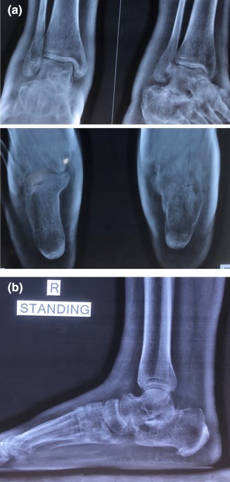 Broken Calcaneus Stock Photos – Free & Royalty-Free Stock Photos from Dreamstime – #79
- x ray heel ap lateral view
- calcaneus lateral view
- harris view calcaneus fracture
 Postoperative lateral radiograph of the calcaneus shows anatomical… | Download Scientific Diagram – #80
 Calcaneus – an overview | ScienceDirect Topics – #81
 Ankle x-rays – Don’t Forget the Bubbles – #82
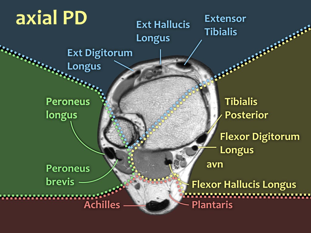 X-ray image of Axial calcaneus (heel) showing achilles tendon rupture and heel fracture Stock Photo | Adobe Stock – #83
 Dual-Position Weight Bearing Cassette & DR Panel Holder – RC Imaging – #84
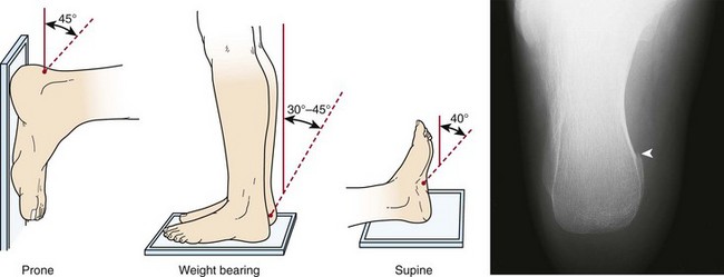 Case Study: Calcaneus fracture – NYSORA – #85
- both heel lateral x ray
- lateral calcaneus x ray positioning
- harris view calcaneus c-arm
 Imaging of the Calcaneus – #86
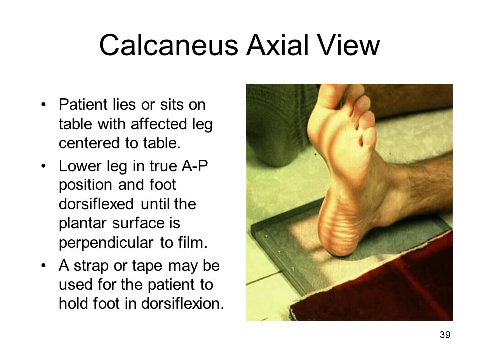 Current management of intra-articular calcaneal fractures | Revista Española de Cirugía Ortopédica y Traumatología (English Edition) – #87
 The Lower Limb – Clark’s Positioning In Radiography – by A. S. Whitley – #88
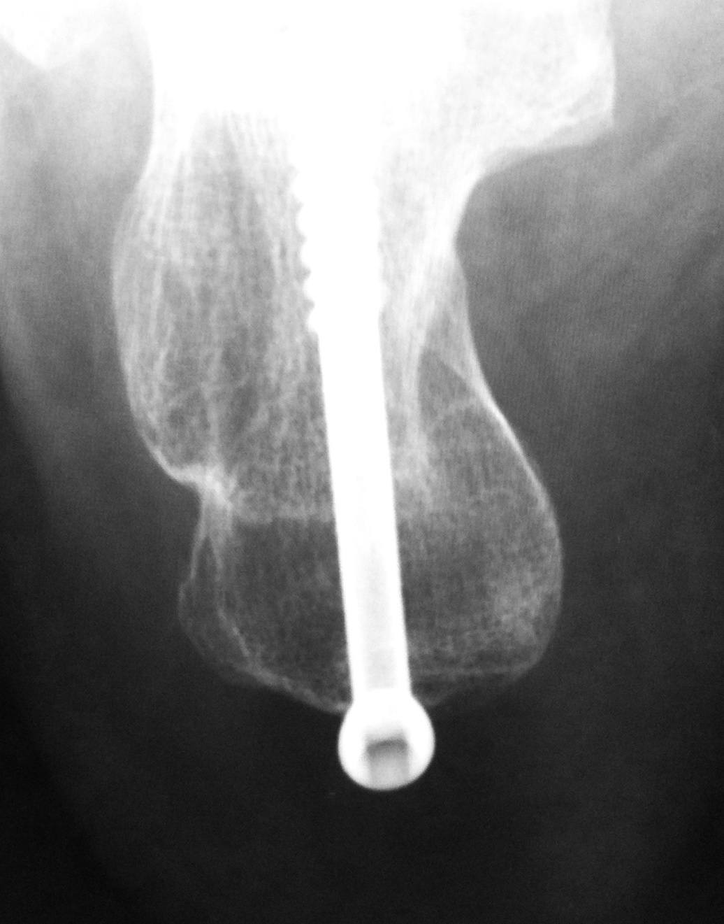 LearningRadiology – Calcaneal, calcaneous, Stress, Fracture – #89
- x ray heel lateral view positioning
- axial dorsoplantar projection calcaneus
- axial calcaneus x ray labeled
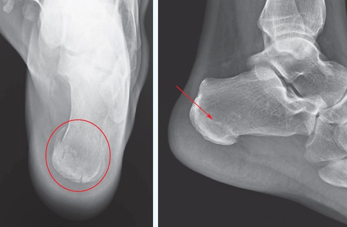 Calcaneus Fractures Workup: Laboratory Studies, Plain Radiography, Computed Tomography – #90
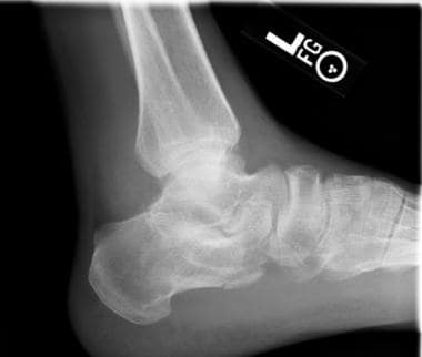 Normal calcaneum series | Radiology Case | Radiopaedia.org – #91
 A new radiographic view of the hindfoot – Ikoma – 2013 – Journal of Foot and Ankle Research – Wiley Online Library – #92
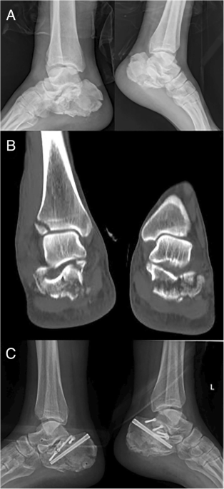 Steinmann pin retractor-assisted reduction with circle plate fixation via sinus tarsi approach for intra-articular calcaneal fractures: a retrospective cohort study | Journal of Orthopaedic Surgery and Research | Full Text – #93
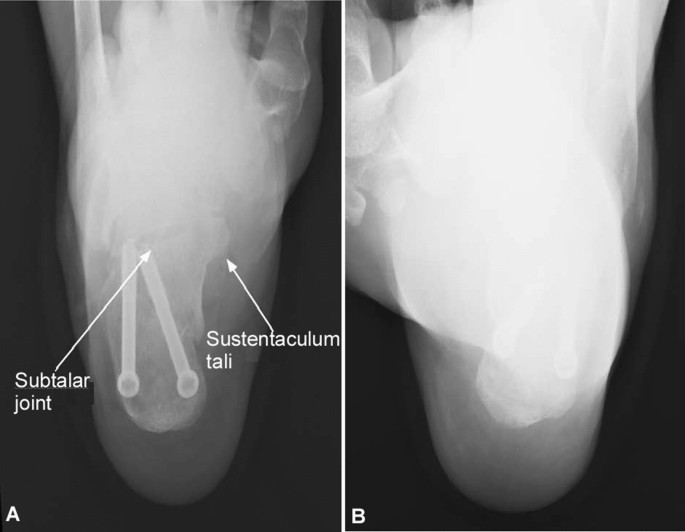 158 Calcaneal Fracture Royalty-Free Photos and Stock Images | Shutterstock – #94
 Tendon and Peritendinous Infections | Radsource – #95
 Orthopaedic Surgery – #96
Posts: heel x ray axial view
Categories: Heels
Author: dienmayquynhon.com.vn
