Details more than 120 both heel lateral x ray latest
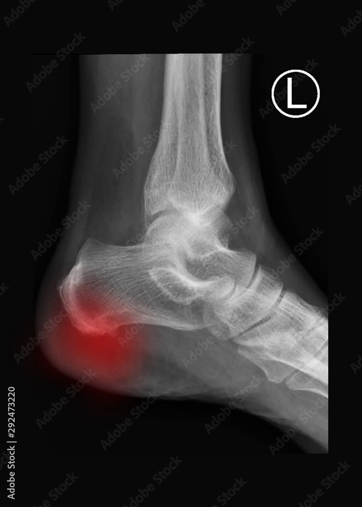
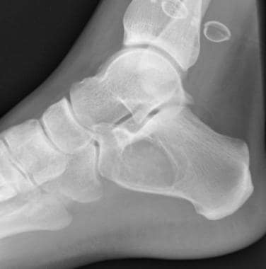
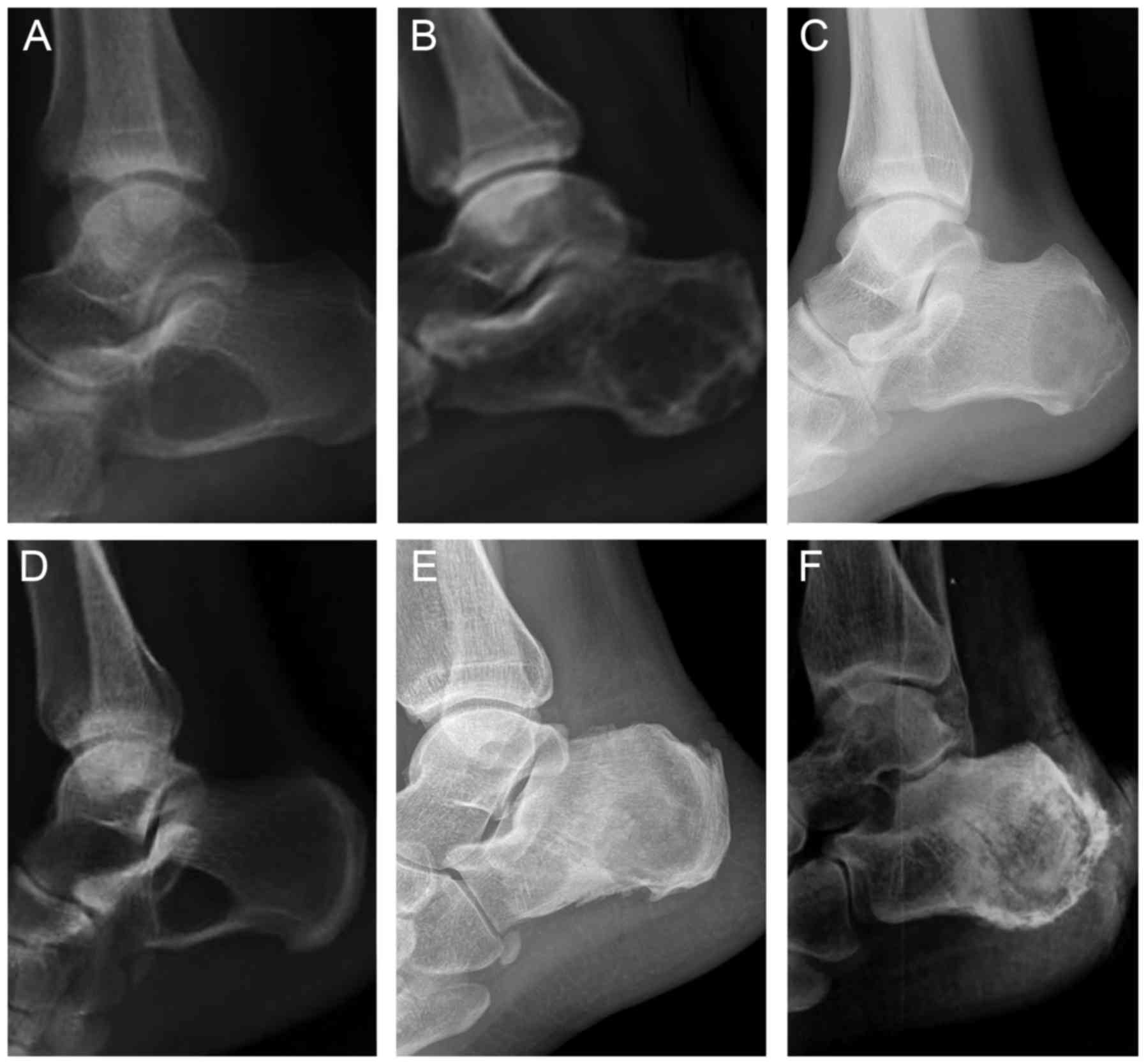
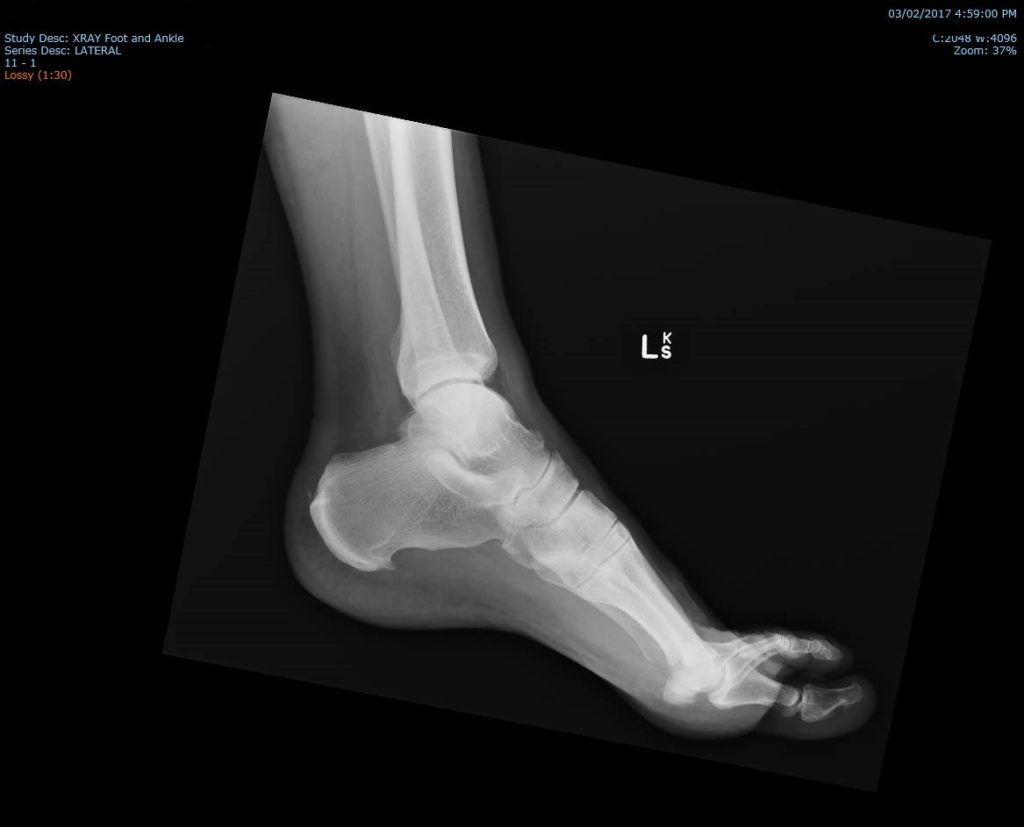
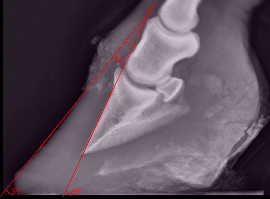
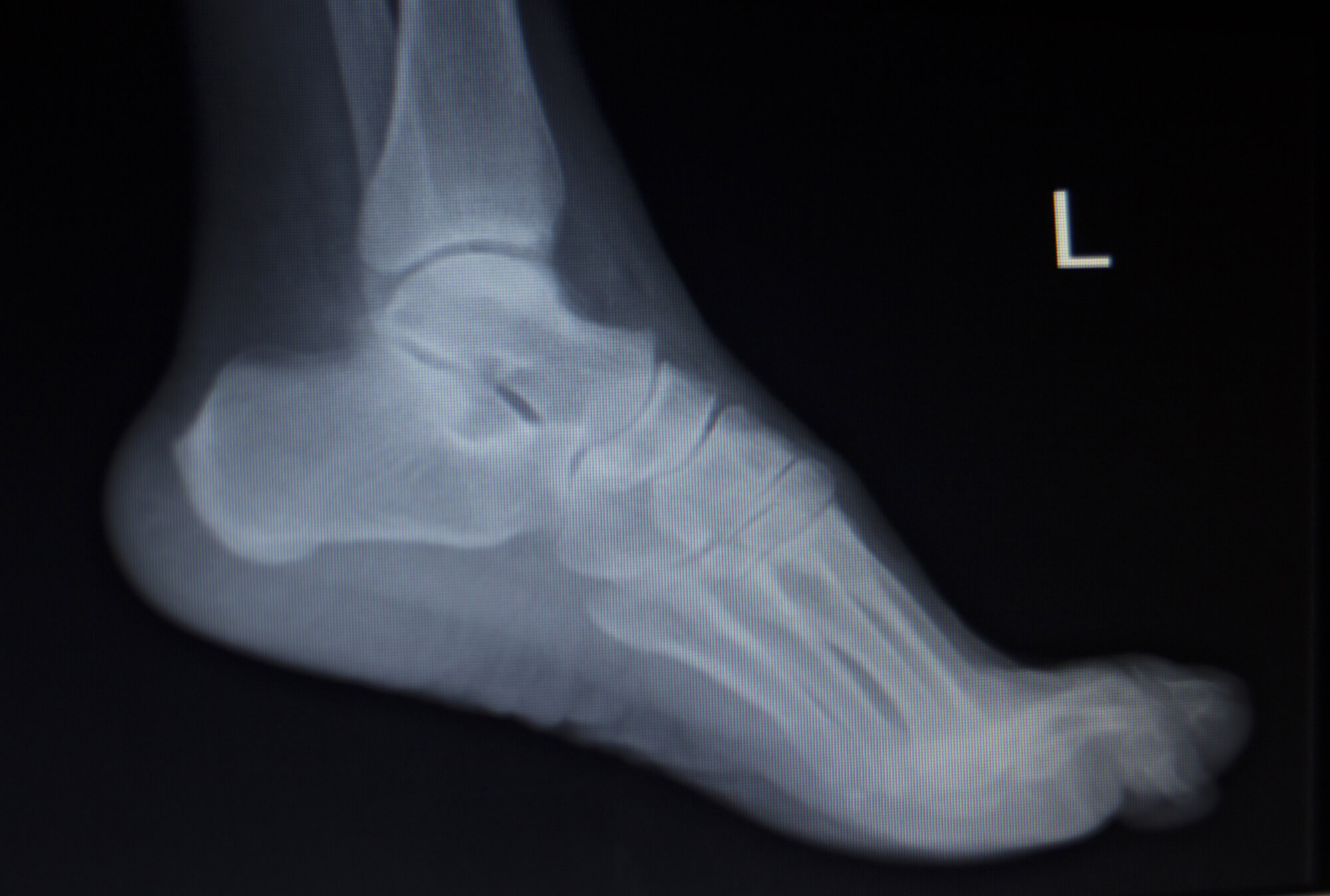
Details images of both heel lateral x ray by website dienmayquynhon.com.vn compilation. X-ray Image of Ankle, Lateral View. Stock Image – Image of scan, metatarsal: 53839907. Distraction arthroplasty in osteoarthritis of the foot and ankle. Radiography techniques of the equine fetlock joint. Calcaneal Fracture-Dislocation With Fracture of the Sustentaculum and Lateral Column: A Unique Injury Pattern – Jeffrey J. Nepple, Ryan M. Putnam, Michael J. Gardner, Craig S. Bartlett, Jeffrey E. Johnson, 2013
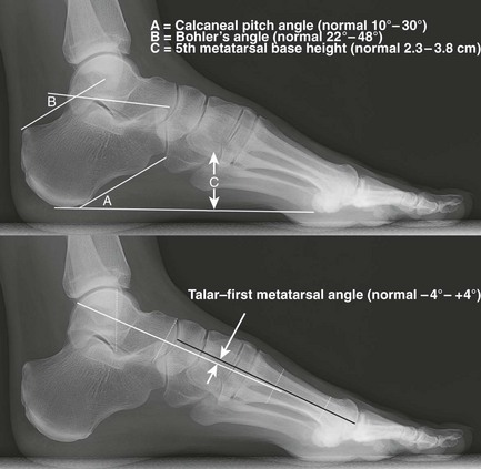
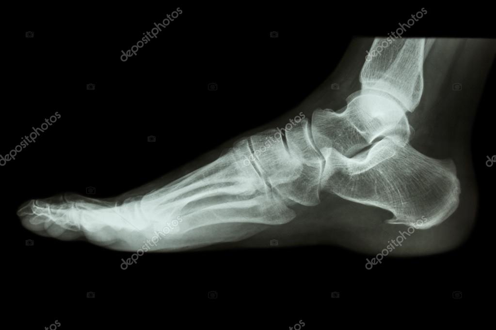
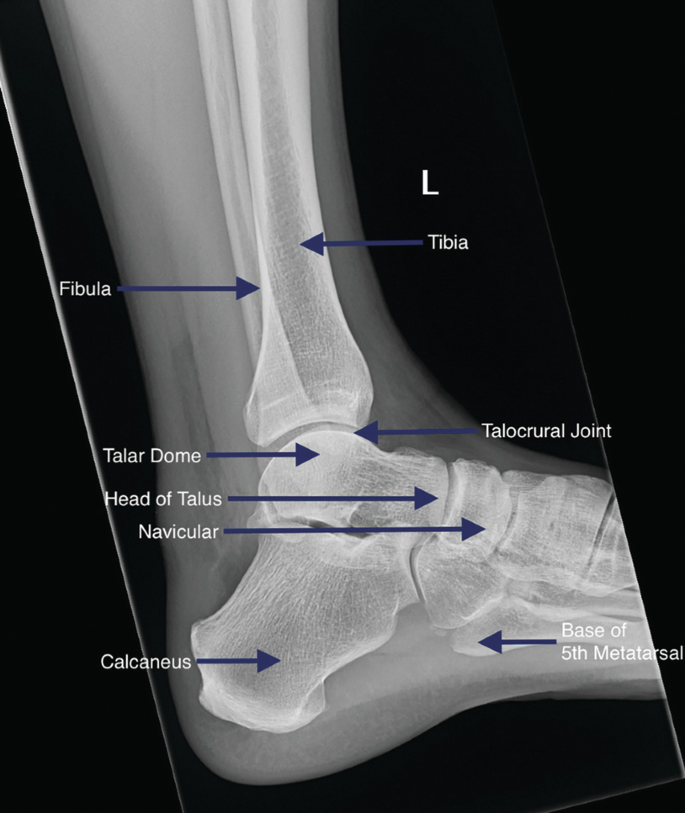
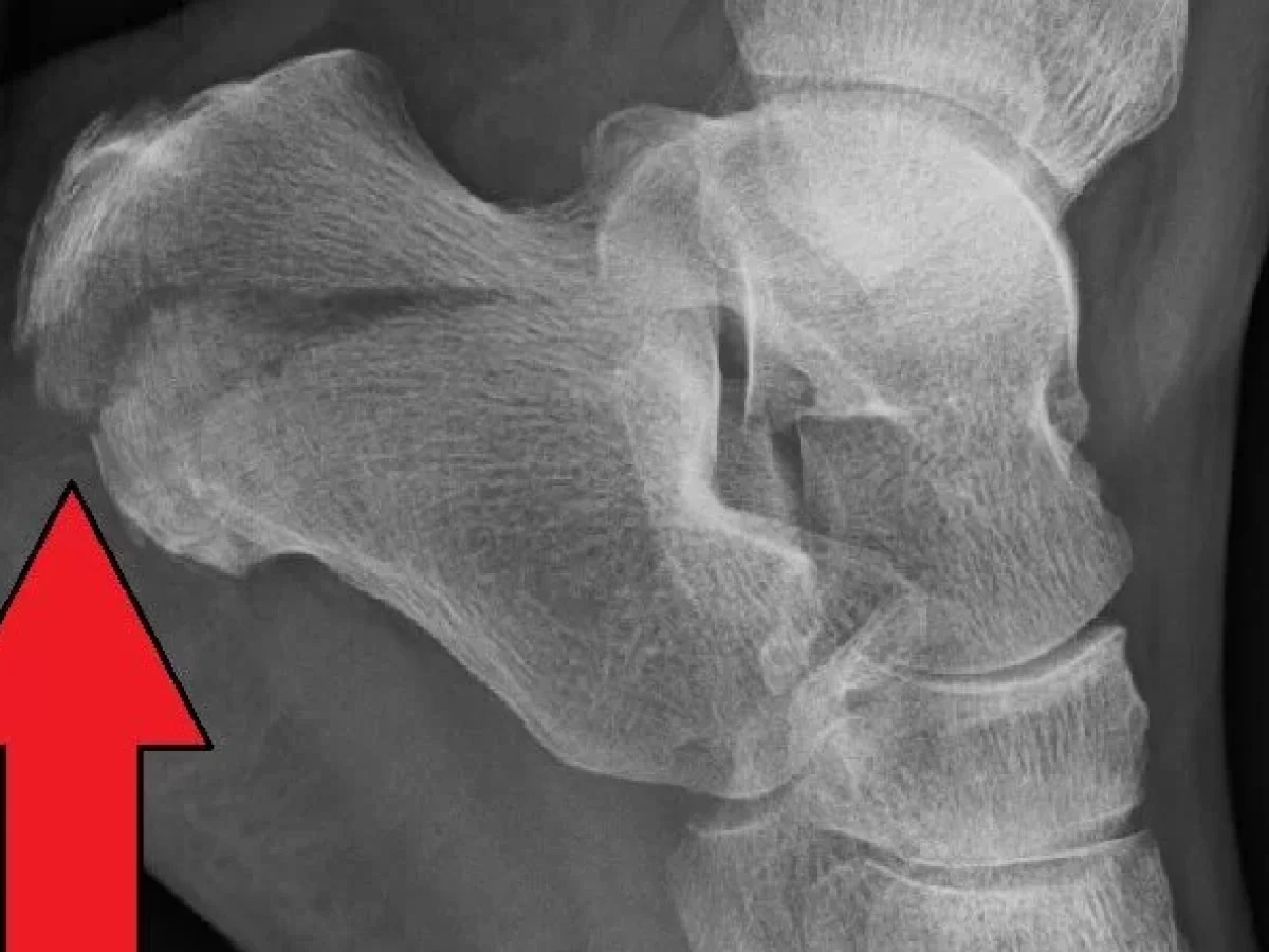 Imaging of the Foot and Ankle | Musculoskeletal Key – #1
- normal lateral foot xray
- ankle x ray
- x ray heel ap lateral view position
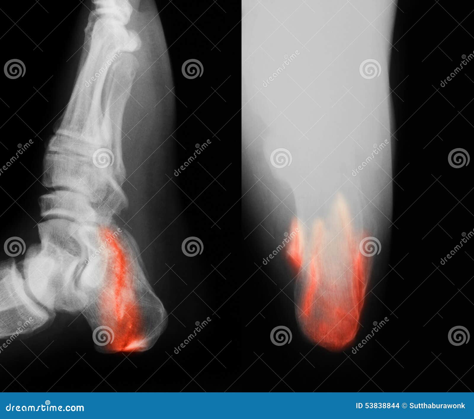
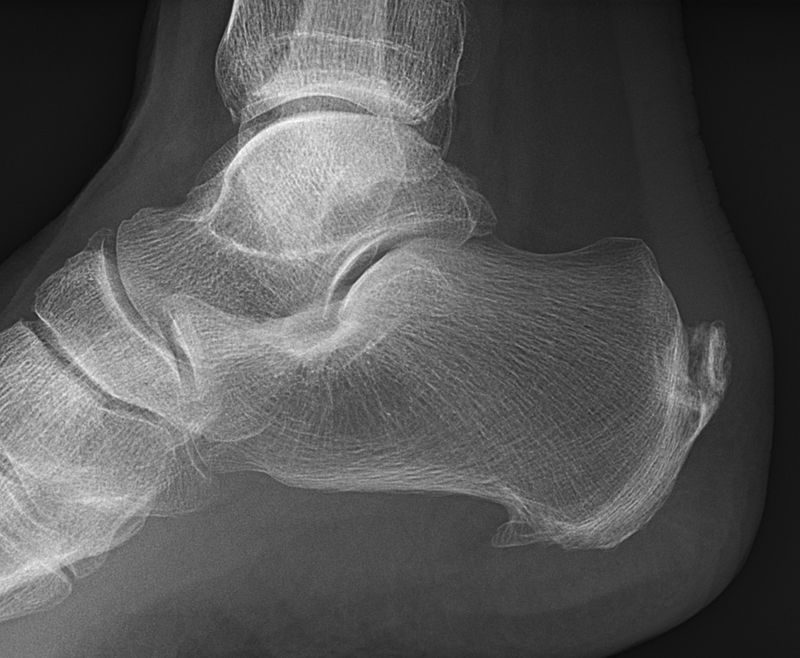 female feet xray radiograph 4965957 Stock Photo at Vecteezy – #2
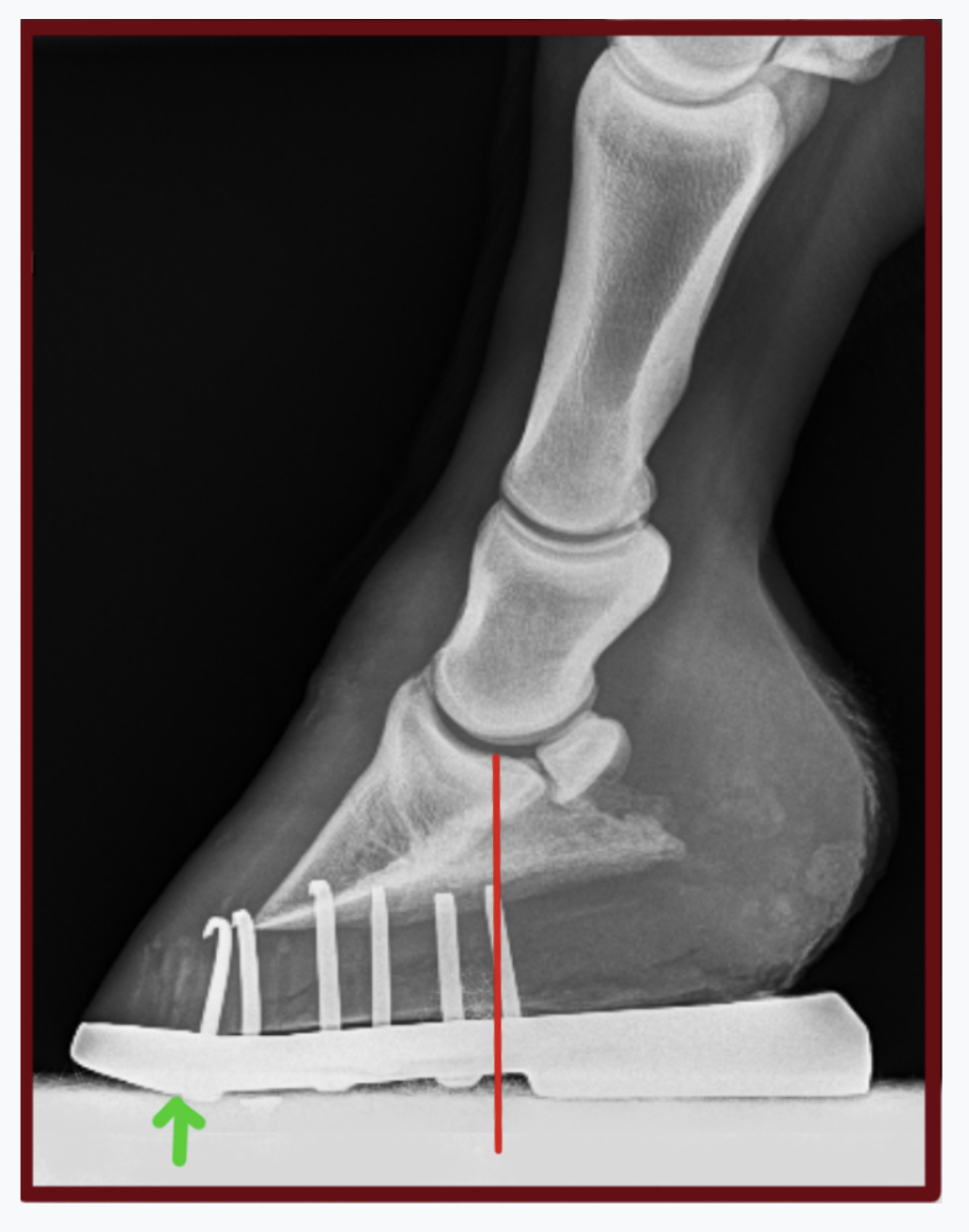
 Xray Image Broken Calcaneusheel Lateral Axial Stock Photo 269956952 | Shutterstock – #3
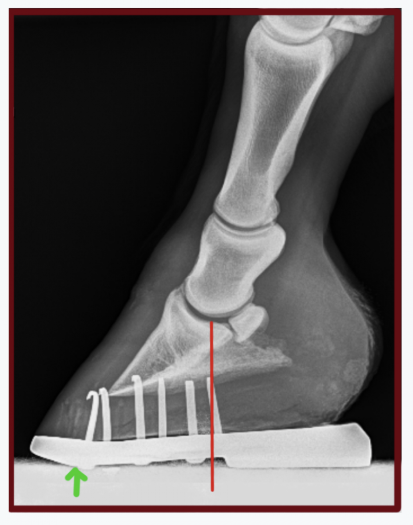
 Flat Feet Explained: Part 2 Non-Surgical Treatment – Scott R. Kilberg DPM – #4
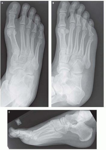 Foot Drop: What It Is, Causes, Symptoms & Treatment – #5
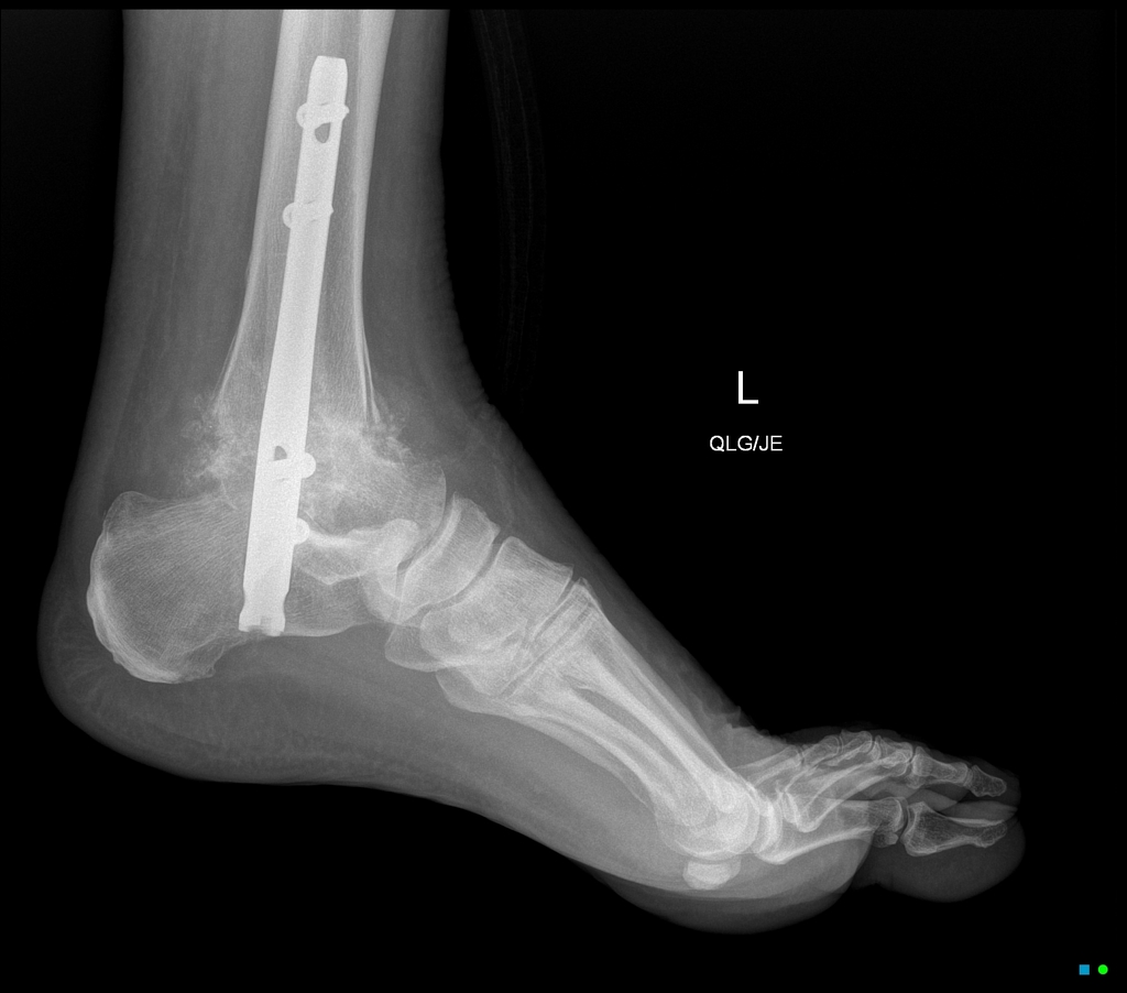 Achillodynia – The Achilles Tendon Pain Syndrome – #6
 Fundraiser for Pete Carlson by Bill Wulff : Pete Carlson’s Recovery Fund – #7
 Why Do I Have Heel Pain After Running? – Atlas Pain Specialists – #8
 Reliability and validity of radiographic measurements in hindfoot varus and valgus. | Semantic Scholar – #9
 MedPix Case – Jones Fracture – #10
 Acupuncture for Tibialis Anterior Pain — Morningside Acupuncture NYC – #11
 X-Ray Both Heel Lat View | Test Price in Delhi | Ganesh Diagnostic – #12
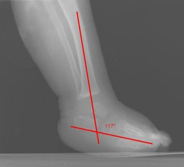 Calcaneus series | Radiology Reference Article | Radiopaedia.org – #13
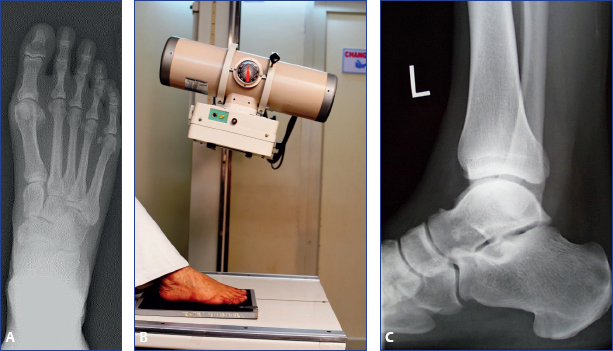
 Distraction arthroplasty in osteoarthritis of the foot and ankle – #14
 Lateral Radiograph of Ankle With Calcaneal Spur. | Download Scientific Diagram – #15
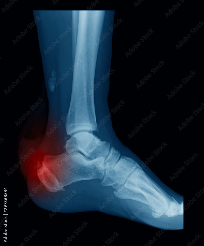
 normal radiography of the ankle joint in the lateral projection Stock Photo | Adobe Stock – #16
 Dr. OMID BANDARCHI on X: “🛑Post fat pad is not visible in normal condition but in children with a distal humeral Fx,can be seen. 🛑Ant fat pad is visible in normal condition,however – #17
 Heel Xray stock image. Image of broken, heel, fracture – 10900219 – #18
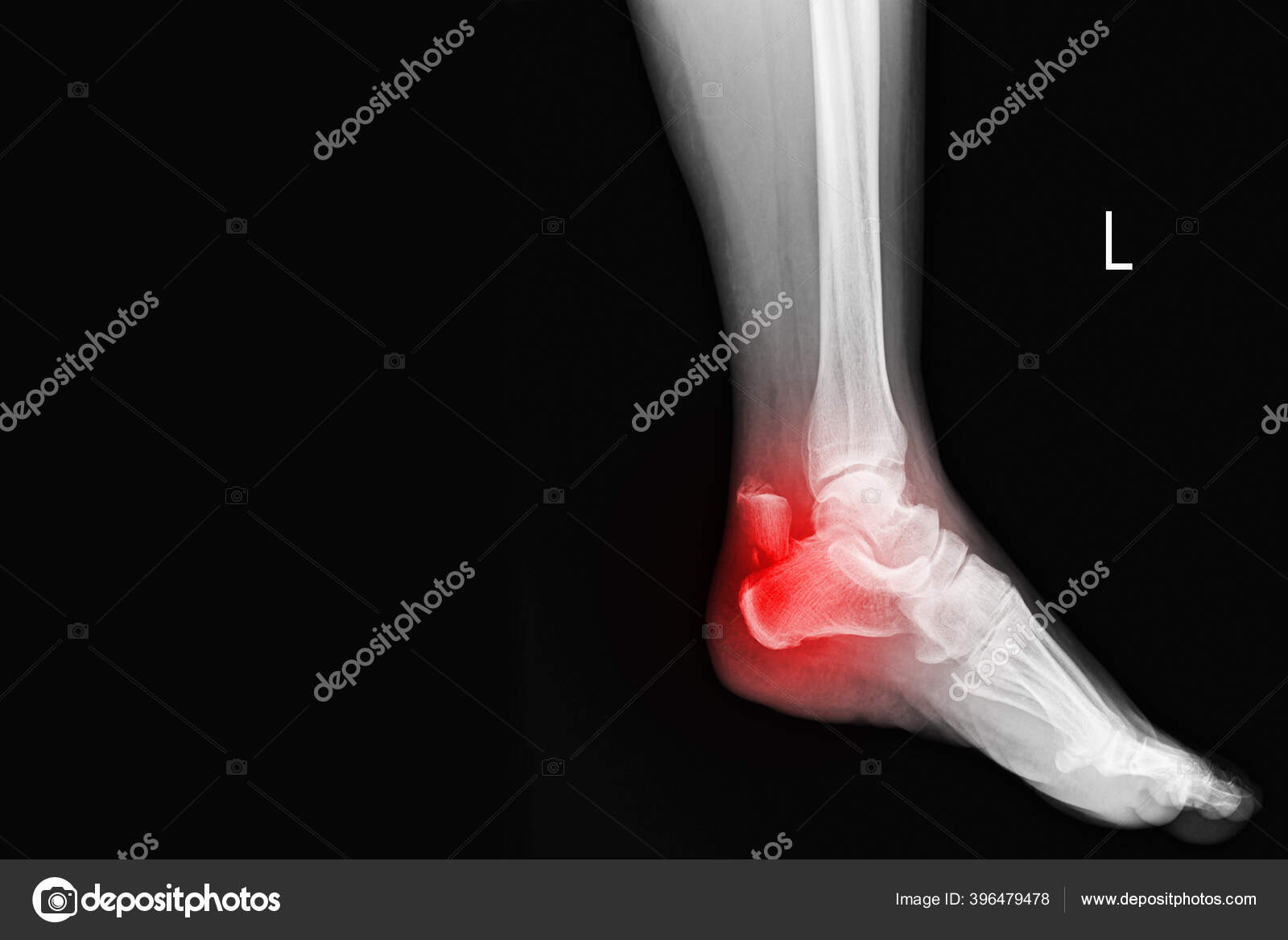 Film Ankle X-ray Radiograph Showing Heel Bone Broken 3 Views Close Fracture Calcaneus . Medical Technology and Healthcare Stock Photo – Image of health, orthopedic: 242419886 – #19
- x ray heel ap view positioning
- heel spur xray vs normal
- achilles heel spur
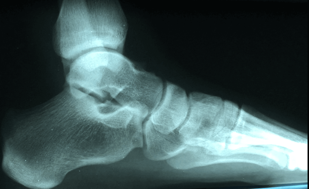 Lateral Ankle Sprain | Complete Physio – #20
 What Is Avulsion Fracture, How To Prevent And Rehabilitate It? — Best Bainbridge Island Physical Therapy Clinic For Pain Relief, Injury Prevention & Rehabilitation – #21
 Before and After – Heel Spur Removal – YouTube – #22
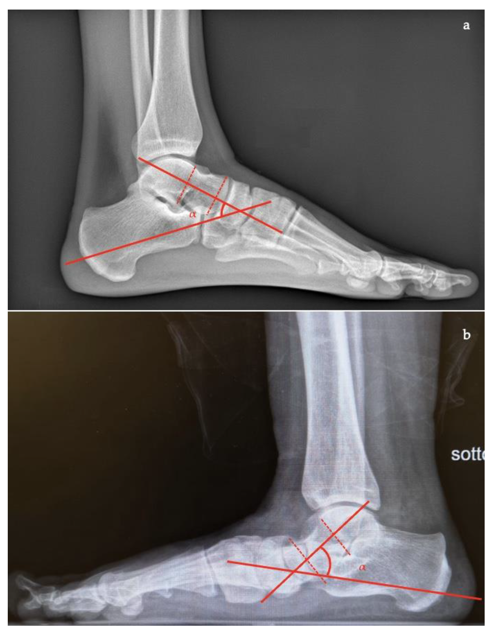 Distal phalanx of left fifth toe | BioDigital Anatomy – #23
 COMMENT YOUR ANSWER 🎯✌️ #neetpg #fmge #inicet | Instagram – #24
 Thank You – Endocrinologist – #25
 Jones Fracture 101: Symptoms, Causes, & Available Treatments – #26
 Lateral Malleolus Fracture Symptoms and Treatment – #27
 Radiology report said normal, but concerned about 4th toe and potential early OA changes : r/xrays – #28
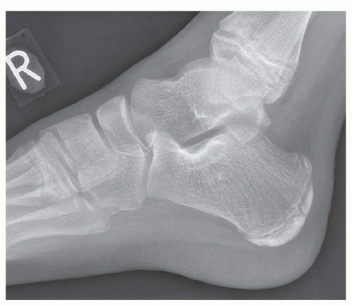 X-ray Image of Ankle, AP and Lateral View. Stock Image – Image of joint, metatarsal: 53839873 – #29
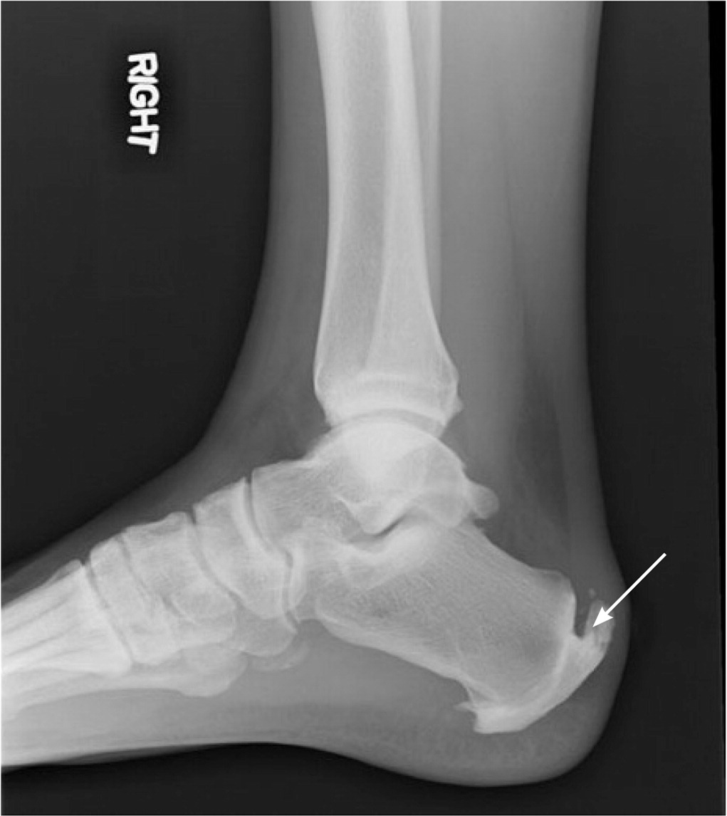 Fractured bone spur – Damien Lafferty – #30
 Tongue-Type Calcaneal Fracture Image 10 Unannotated – JETem – #31
 Ultrasound and Radiographic Abnormalities in a Patient With Chronic Severe Acromegaly | Reumatología Clínica – #32
 JCM | Free Full-Text | Predictive Radiographic Values for Foot Ulceration in Persons with Charcot Foot Divided by Lateral or Medial Midfoot Deformity – #33
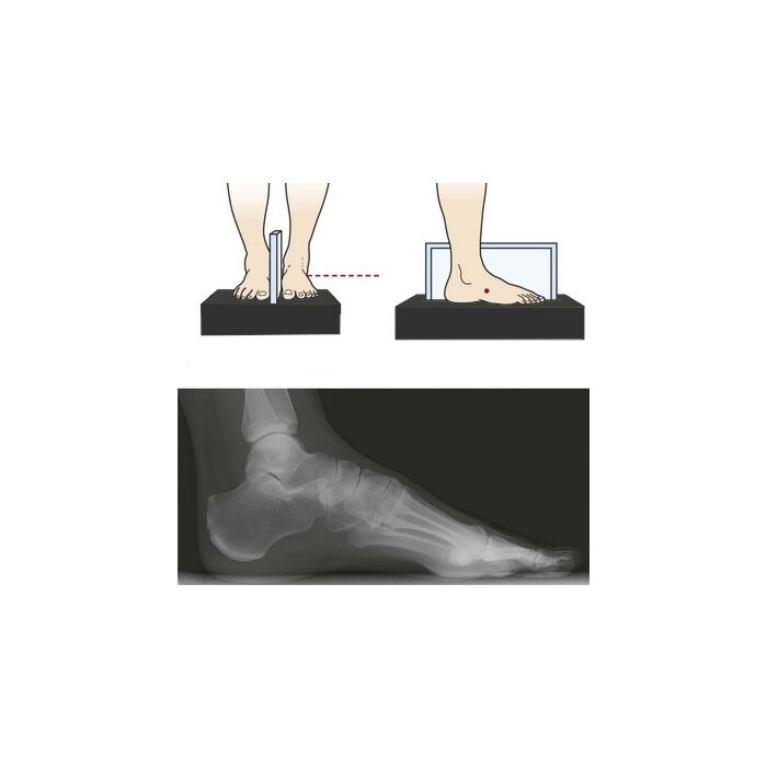 The Role of Periarticular Osteotomies in Total Ankle Replacement | SpringerLink – #34
 X-ray Image Of Broken Calcaneus(heel), Lateral View. Stock Photo, Picture and Royalty Free Image. Image 38810461. – #35
 File:X-ray of a normal foot of a 12 year old male – lateral.jpg – Wikimedia Commons – #36
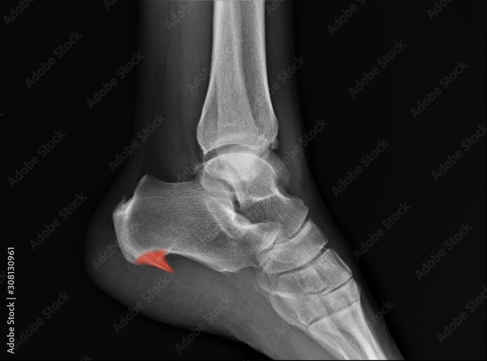 Understanding x-rays – The Laminitis Site – #37
 Figure 4 from Adult-Acquired Flatfoot Deformity: Etiology, Diagnosis, and Management | Semantic Scholar – #38
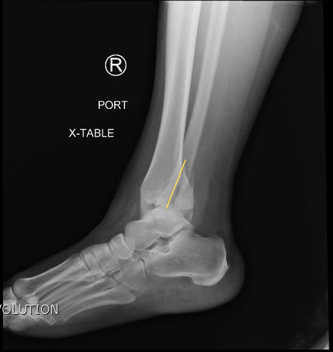 Does Your Foot Hurt Here?: The Outside of the Foot – #39
 Haglund deformity of the posterior heel – #40
 Premium Photo | A broken ankle is shown with a bone in the middle. – #41
 X-ray Image Of Ankle Joint , Lateral View. Show Ankle Fracture. Stock Photo, Picture and Royalty Free Image. Image 39882426. – #42
 Diagnosing Knee Hyperextension | Sports-health – #43
 Foot Xray High-Res Stock Photo – Getty Images – #44
 DBMCI Egurukul – Question of the day #latestpatternquestion A 44 year old male, policeman by profession , reported heel pain for approximately 1 year and tenderness at the calcaneal tuberosity. The pain – #45
 From the EMDaily Archives: What’s the Diagnosis? By Dr. Rebecca Fieles – – #46
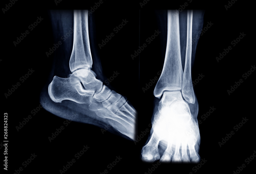 Bone Spurs on Your Foot – What to Do in Scottsdale – Arizona Foot Doctors – #47
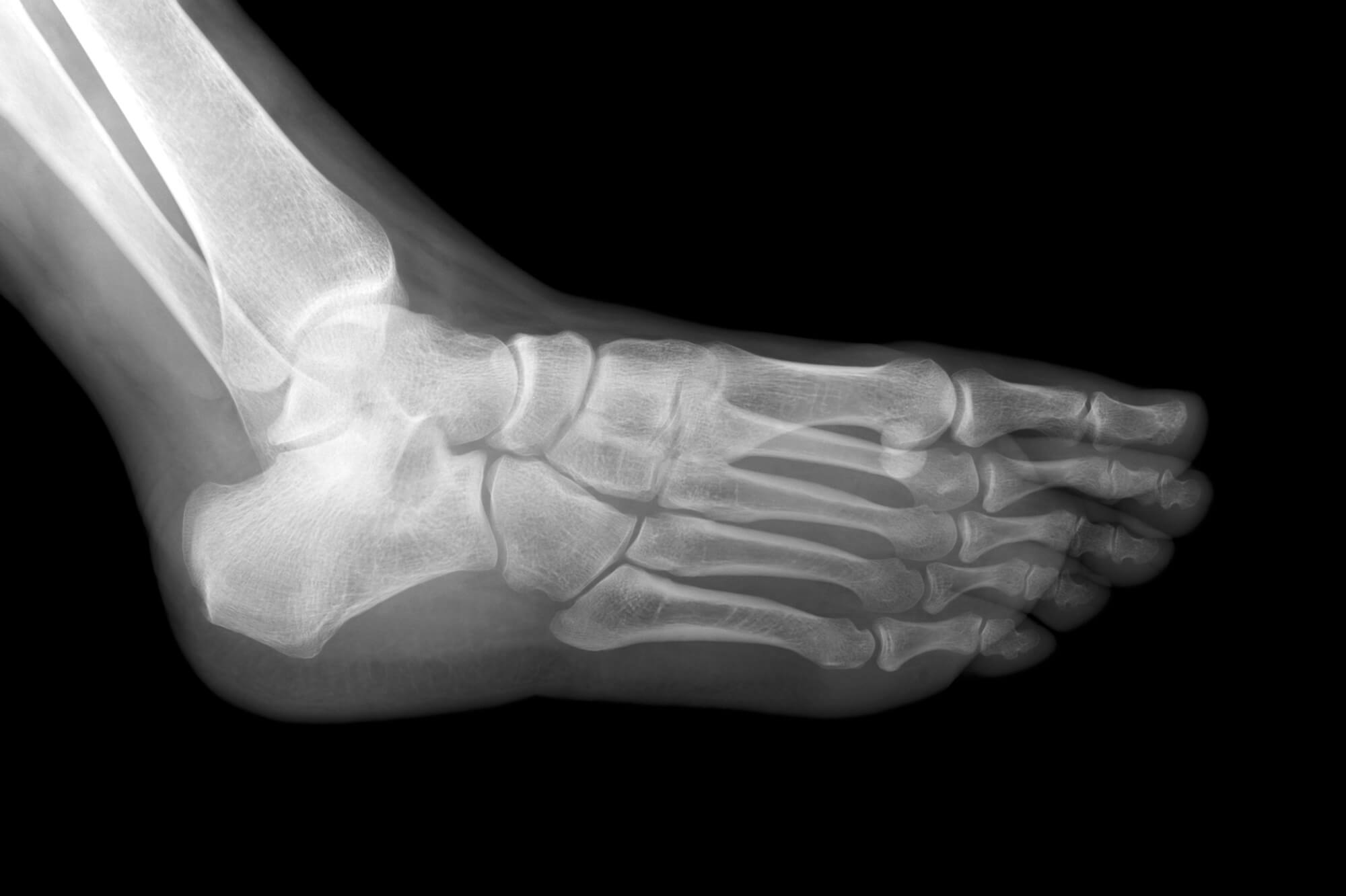 OrthoDx: Heel Pain in 12-Year-Old Boy – Clinical Advisor – #48
- plantar fasciitis x ray vs normal
- x ray heel lateral view positioning
- normal heel x ray
 MedPix Topic – Melorheostosis – #49
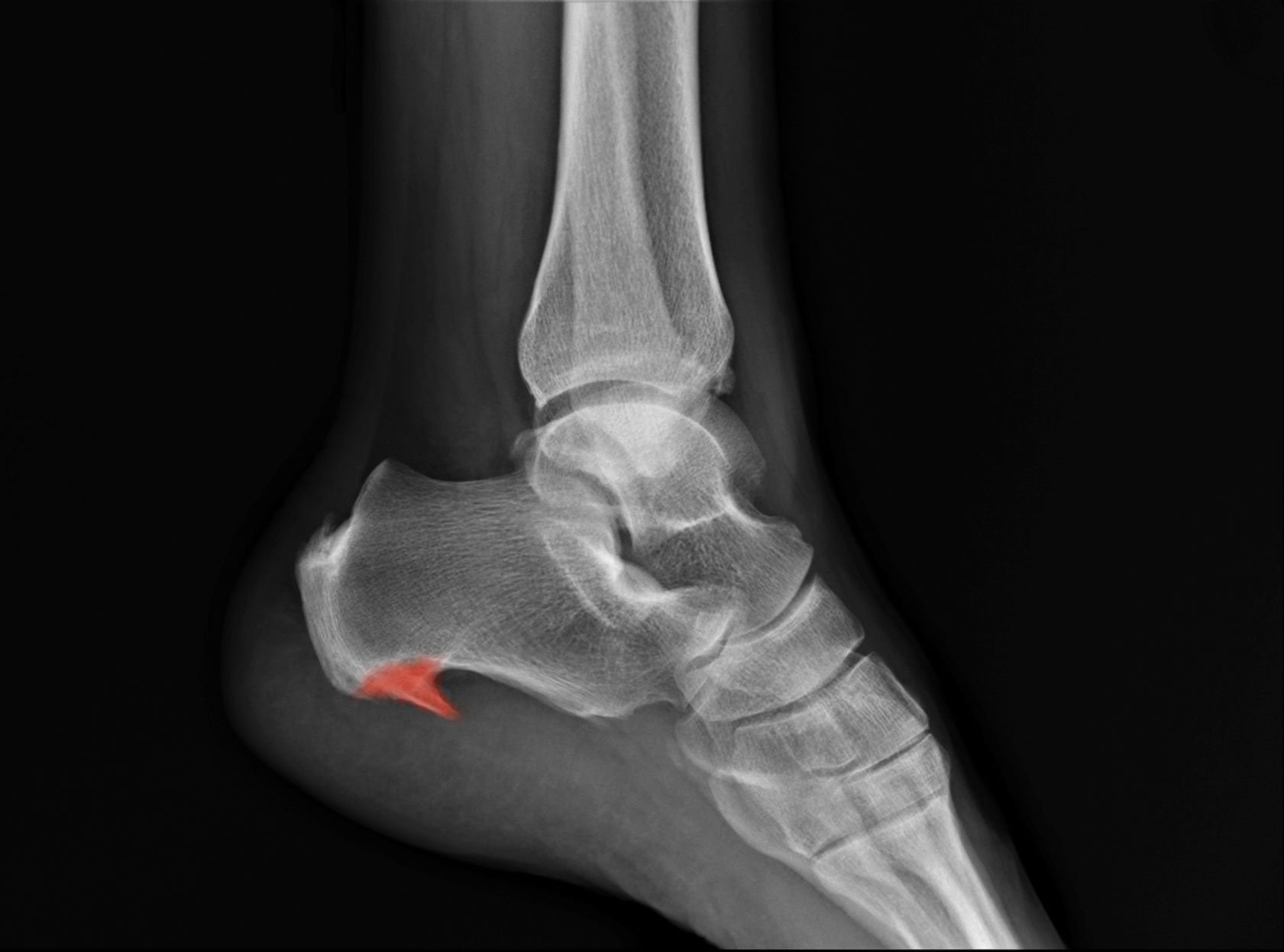 Avulsion fracture of the calcaneus | CMAJ – #50
 X-ray Both Foot Ap. Image & Photo (Free Trial) | Bigstock – #51
 Ray Left Knee Lateral Showing Kneecap Normal Fracture Red Color Stock Photo by ©Richmanphoto 218602808 – #52
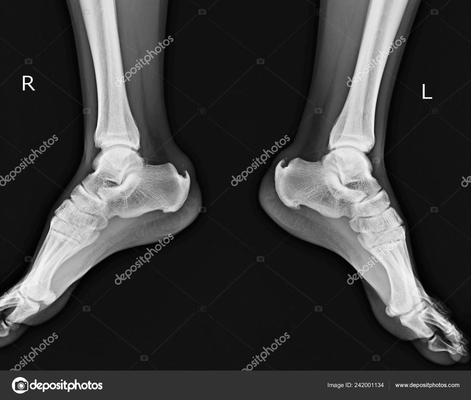 Radiology Quiz 75155 | Radiopaedia.org – #53
 X-Ray Bilateral Heel AP View and Lateral | Test Price in Delhi | Ganesh Diagnostic – #54
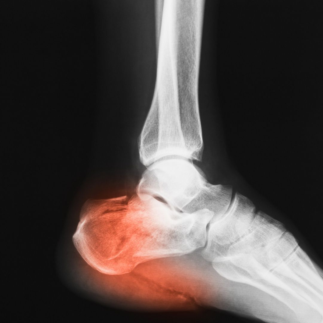 A) Lateral X-ray of calcaneus, obvious displacement of calcaneal… | Download Scientific Diagram – #55
 Assessing Heel Pain | Diagnostic Ultrasound of the Foot and Ankle – #56
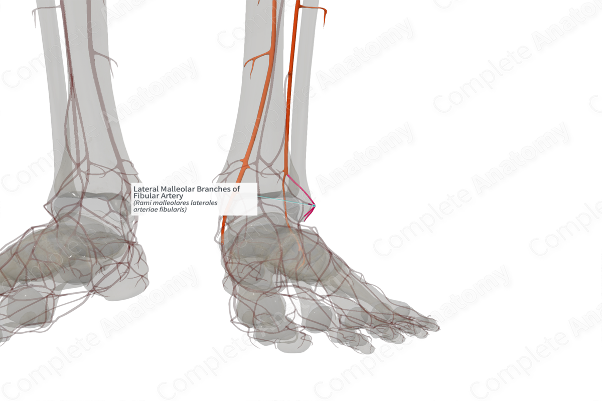 Foot xray hi-res stock photography and images – Alamy – #57
 John Hawks on X: “The human foot and ankle together are a remarkable structure. 26 bones plus sesamoids, more than 30 joints, and more than 100 ligaments, tendons, and muscles. Our evolution – #58
 Heel Pad Sign – #59
 When there is misalignment astragalus heel, around ten or twelve years of age, it can be corrected by surgery. | Radiology, Pes planus, Brainstorming – #60
 Flat Feet – The Center for Advanced Orthopedics – #61
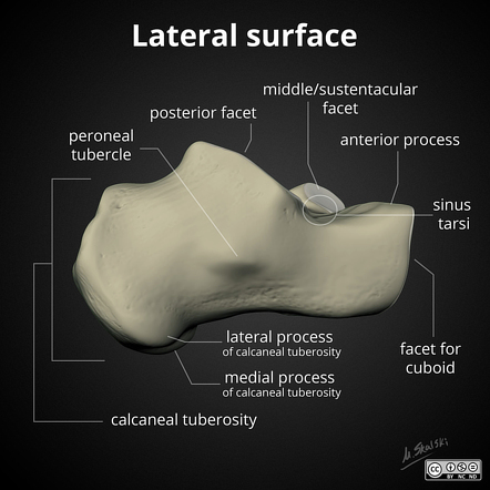 The Charcot foot: a pictorial review | Insights into Imaging | Full Text – #62
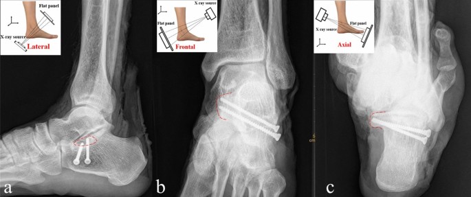 Subtalar Joint Fusion — Chicago Foot & Ankle Orthopaedic Surgeons – #63
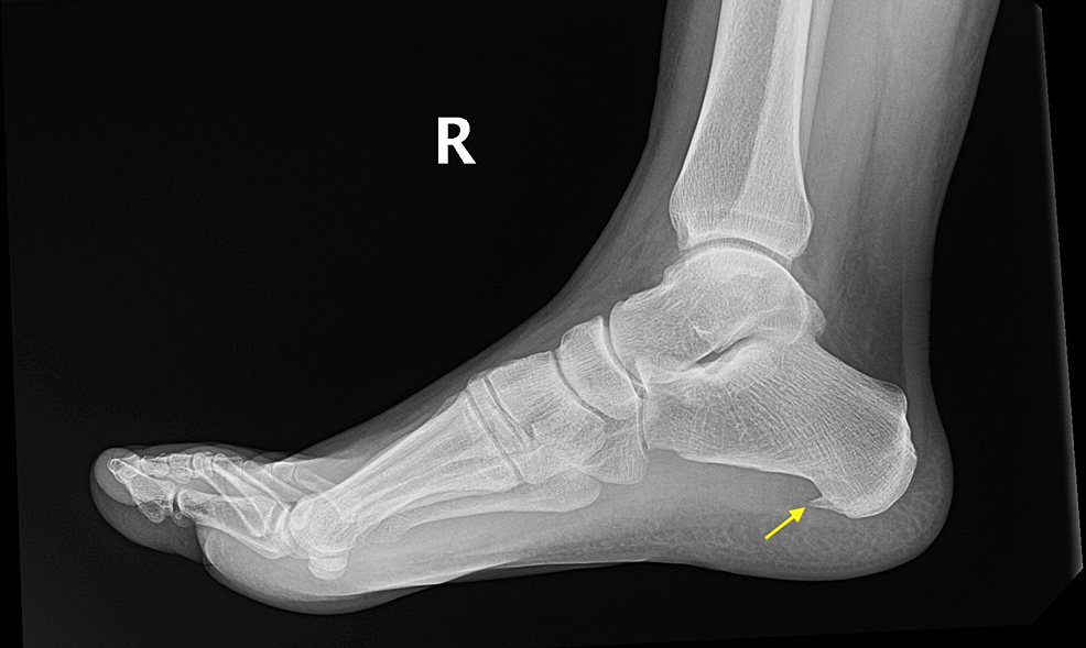 X-Ankle – #64
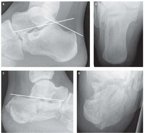 Lateral x-rays of both feet. Note the different thicknesses of the heel… | Download Scientific Diagram – #65
 EMRad: Radiologic Approach to the Traumatic Ankle – #66
- plantar fasciitis normal heel xray
- heel lateral position
- röntgenbild ferse gebrochen
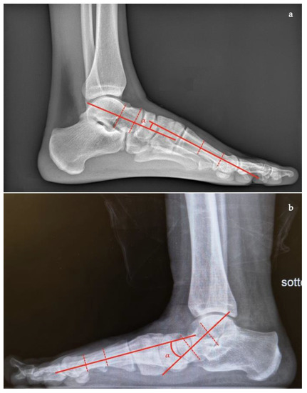 Diagnosis of a Radiographically Occult Calcaneus Fracture With Bedside Sonography – Augustin-Coley – 2016 – Journal of Ultrasound in Medicine – Wiley Online Library – #67
 Exostosis: Causes, types, and treatment – #68
 X-ray Left Heel Lateral/Axial | Test Price in Delhi | Ganesh Diagnostic – #69
 Film X-ray Normal Human’s Foot Lateral Stock Photo, Picture and Royalty Free Image. Image 33990618. – #70
 Painful Heel Spur Management in Singapore | Podiatry Clinic – #71
 Xray Image Of Foot Lateral View Stock Photo – Download Image Now – Human Foot, Spur, Calcaneus – iStock – #72
 1 in 3 Unstable Ankles Has Cartilage Damage – Regenexx – #73
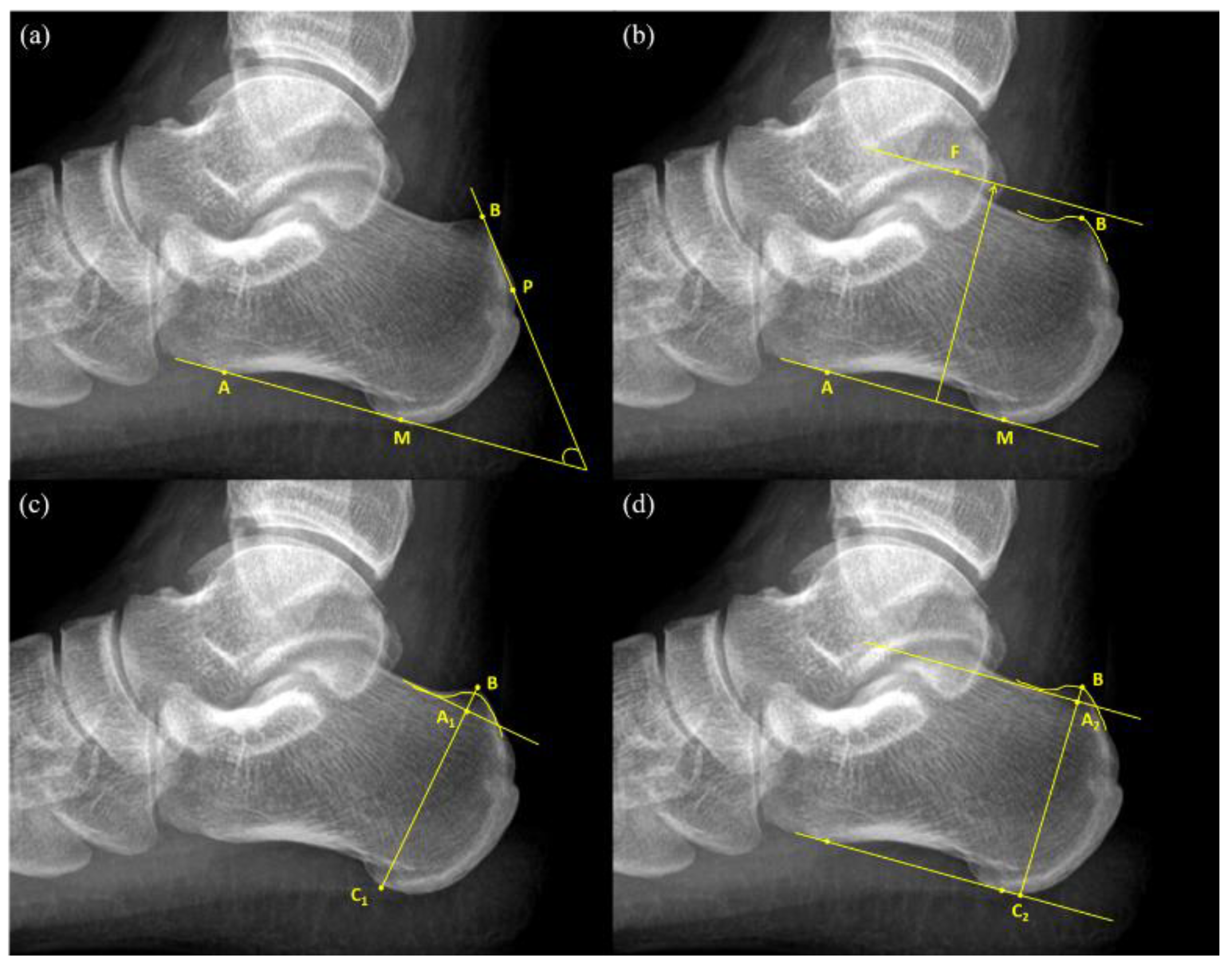
 Calcaneal Fractures and Böhler’s Angle – JETem – #74
 X ray heel or calcanius axial & lat view ( ep -11) |Bangla tutorial review | positioning of heel. – YouTube – #75
 x ray ankle ap lateral Stock Photo | Adobe Stock – #76
 CALCANEUS LATERAL POSITIONING HINDI | X RAY POSITIONING FOR RADIOGRAPHERS | DOCTOR INSIDE – YouTube – #77
- both heel ap x ray
- foot x ray
- healthy normal heel xray
 Metatarsal Head Osteotomies For Bunion Correction – #78
![PDF] Radiographic study of Sever's disease | Semantic Scholar PDF] Radiographic study of Sever's disease | Semantic Scholar](https://assets.cureus.com/uploads/figure/file/709987/article_river_953cf31032d011eebd1fe77155cffff5-Figure-1-kopyasi.png) PDF] Radiographic study of Sever’s disease | Semantic Scholar – #79
PDF] Radiographic study of Sever’s disease | Semantic Scholar – #79
 Calcaneal Spurs, X-ray #2 Photograph by Living Art Enterprises – Pixels – #80
 Xray Image Broken Heel Lateral View Stock Photo 274974218 | Shutterstock – #81
- x ray calcaneus lateral view
- normal heel x ray vs heel spur
- calcaneus lateral view position
![Extensor Digitorum Longus - Download Free 3D model by Mary Orczykowski (@anatomary) [7b539aa] Extensor Digitorum Longus - Download Free 3D model by Mary Orczykowski (@anatomary) [7b539aa]](https://i.pinimg.com/474x/fd/44/cb/fd44cb5ded618c14ec40de938cb7ca5d.jpg) Extensor Digitorum Longus – Download Free 3D model by Mary Orczykowski (@anatomary) [7b539aa] – #82
Extensor Digitorum Longus – Download Free 3D model by Mary Orczykowski (@anatomary) [7b539aa] – #82
 Frontiers | Case Report: An Unusual Case of Acute Lower Limb Ischemia as Precursor of the Asherson’s Syndrome – #83
 Film X-ray of Child S Foot ( Side View ) ( Lateral ) Stock Image – Image of heel, lateral: 58744591 – #84
 Xray Of The Heel After Calcaneus Fracture Stock Photo – Download Image Now – Ankle, Prosthetic Equipment, Repairing – iStock – #85
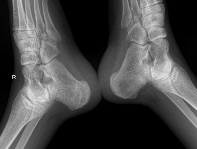
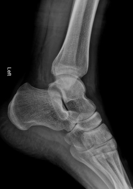
 Lateral view of the plain radiograph of the foot (right and left). | Download Scientific Diagram – #86
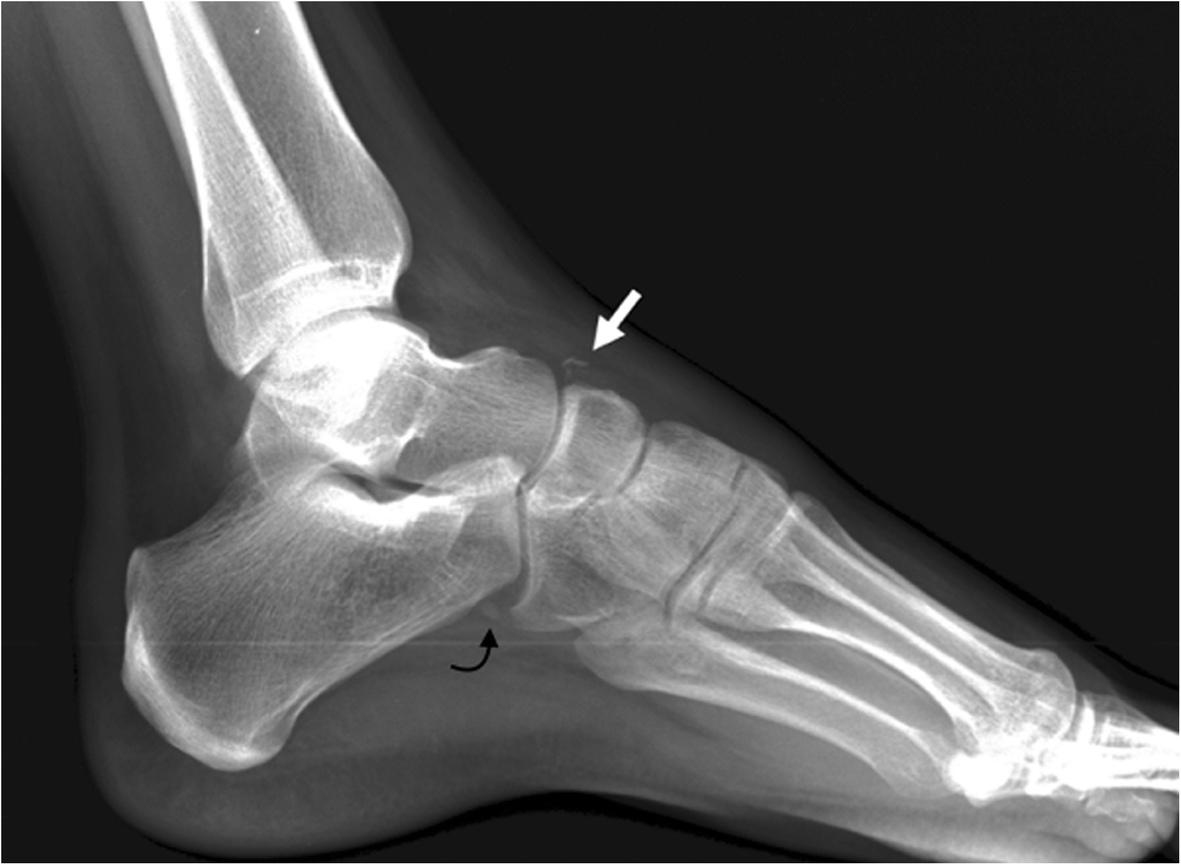
 Iselins Disease: Causes, Symptoms & Treatment – #87
 RiT radiology: Medial Epicondyle Fracture of the Humerus – #88
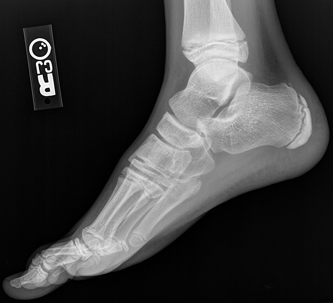 X Ray Normal Foot Lateral People Background Medicine Photo And Picture For Free Download – Pngtree – #89
 Smart implants: surgical nail for monitoring bone healing – TTP – #90
 Systematic Way to Read Foot Xrays – Ortho Conditioning – #91
 Implant zaps the inside of the knee joint to stimulate the growth of cartilage | Daily Mail Online – #92
 Chronic Lateral Ankle Pain: Causes and Treatment Option – #93
 The bends and flexures of forearm and elbow x-ray positioning | AuntMinnie – #94
 Plantar Fasciitis – Union Spine – #95
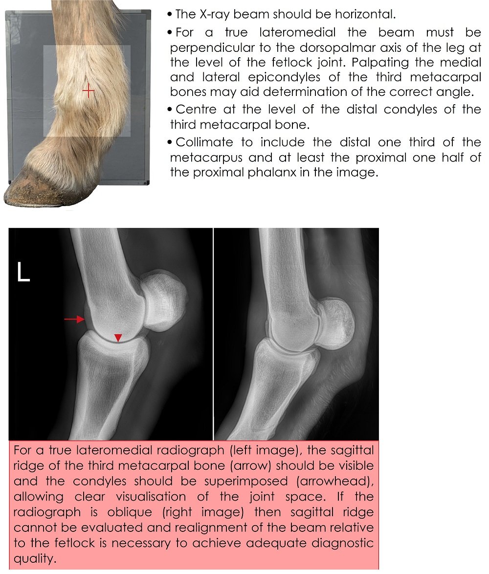
 Radiography techniques of the equine fetlock joint – #96
 Sprains: Types, Symptoms & Treatment – #97
 AP, Lateral and Axial views of the ankle joint showing calcaneal… | Download Scientific Diagram – #98
 Dr. OMID BANDARCHI on X: “Syndesmotic injury may be difficult to Dx,& radiological evaluation is very important. It worths being familiar with normal tibiofibular syndesmosis parameters that help us in diagnosing injuries – #99
 459 Broken Calcaneus Royalty-Free Images, Stock Photos & Pictures | Shutterstock – #100
 Foot and Heel | Radiology Key – #101
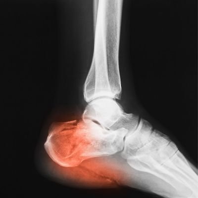
 3,100+ Heel Xray Stock Photos, Pictures & Royalty-Free Images – iStock – #102
 Fractured Heel, X-ray – Stock Image – C027/2614 – Science Photo Library – #103
- heel x ray position
- foot lateral view positioning
- lateral calcaneus x ray positioning
 Heel Pain Series Week 1: Anatomy – Sports and Structural Podiatry – Maroochydore – #104
 Look at the X-Ray … broken Talus? : r/Orthopedics – #105
 normal foot x-ray of a 59 year old woman, Stock Photo, Picture And Rights Managed Image. Pic. CUR-IS09AF27D | agefotostock – #106
 80+ Human Foot Screw X Ray Human Bone Stock Photos, Pictures & Royalty-Free Images – iStock – #107
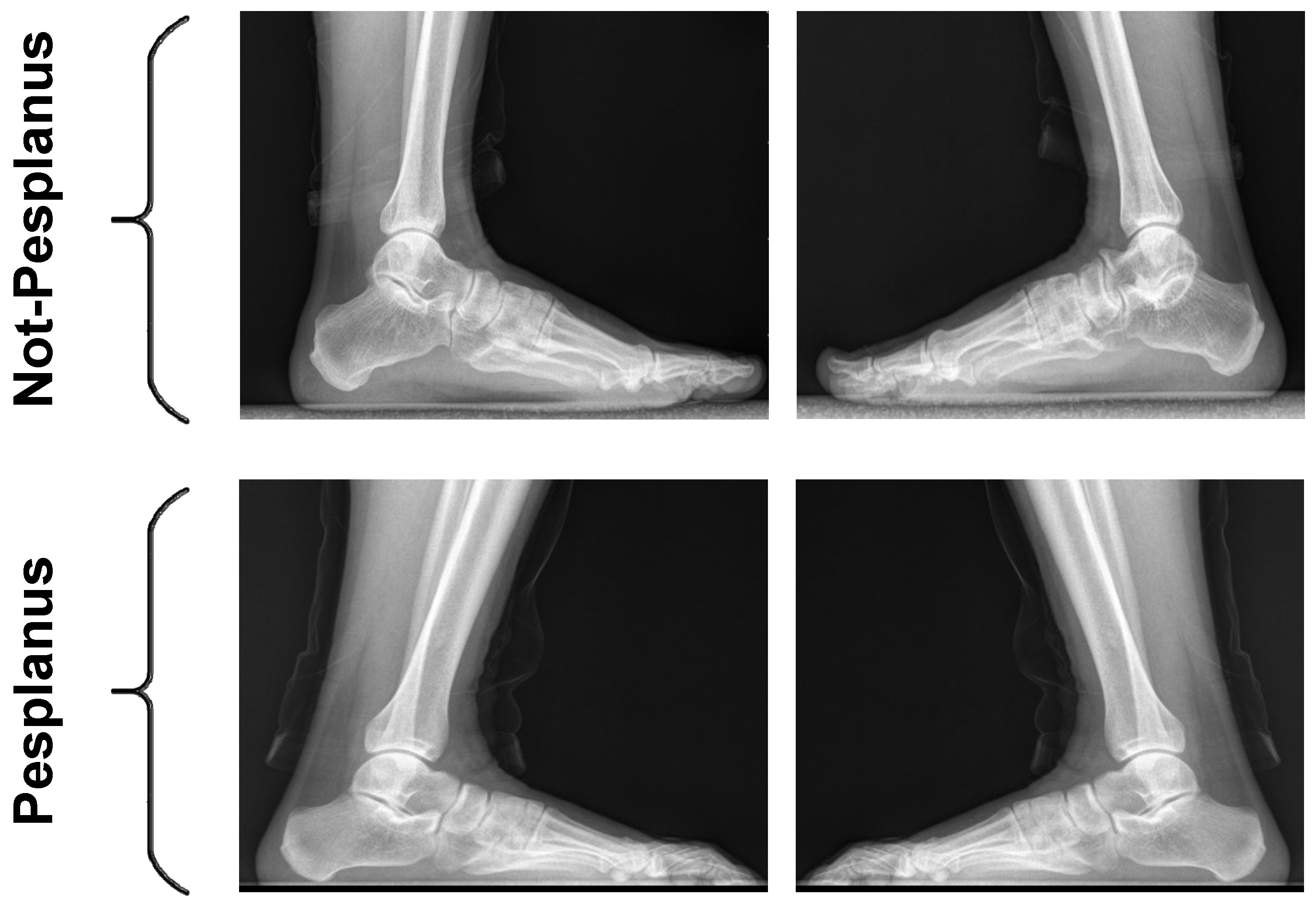
 Film x-ray normal human’s foot lateral Stock Photo | Adobe Stock – #108
 Primary tumours of the calcaneus (Review) – #109
 Medical Science Monitor | Static and Dynamic Plantar Pressure Distribution in 94 Patients with Different Stages of Unilateral Knee Osteoarthritis Using the Footscan® Platform System: An Observational Study – Article abstract #938485 – #110
 Nail in Foot, X-ray | Stock Image – Science Source Images – #111
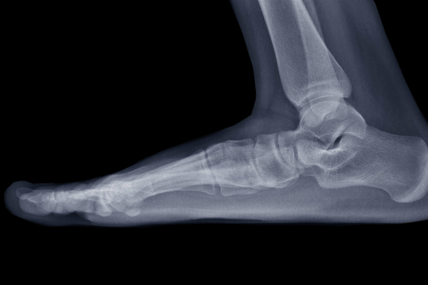 Lateral Right Foot X-ray Showing Destruction of the Posteroinferior Aspect of the Calcaneus Stock Image – Image of finger, defect: 206252641 – #112
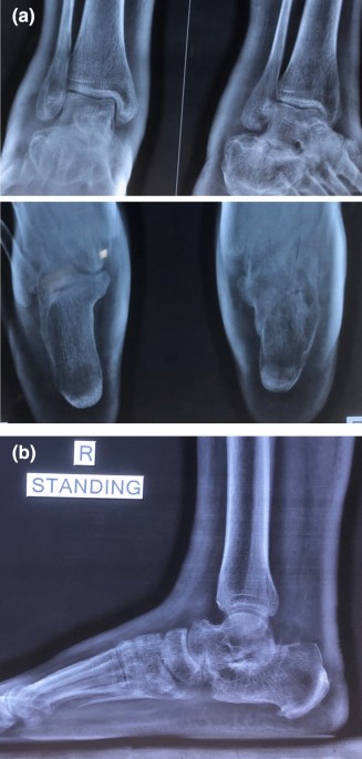 Dr. Young Talks About Lateral Column Pain and Lateral Overload – #113
 Orthobullets on Instagram: “Can you answer our FREE Question of the Day? A 17-year-old female presents with foot pain associated with footwear. A current lateral radiograph of the affected foot is depicted – #114
- heel ap position
- heel spur x ray
- heel spur removal
 Troy J. Boffeli DPM, FACFAS, Tyler K. Sorensen DPM, and Collin G. Messerly, DPM Regions Hospital / HealthPartners Institute for – #115
 Film Ankle Xray Radiograph Showing Heel Stock Photo 1490402393 | Shutterstock – #116
 Dr. OMID BANDARCHI on X: “🛑A Pilon Fracture (=tibial plafond Fx) is any Fx of distal tibia which involves the articular surface of tibia (tibial plafond). Associated with comminution,intra articular extension,..etc. ✴️2 – #117
 images.squarespace-cdn.com/content/v1/5f538bdcabe4… – #118
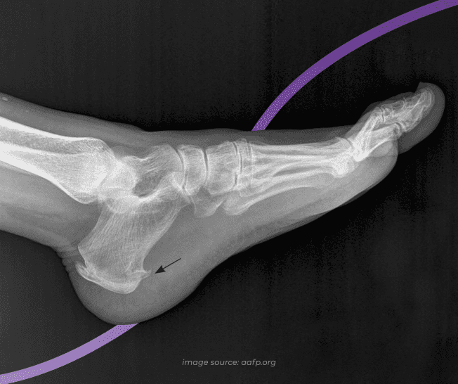 Knee X-Rays and Detecting Abnormalities – #119
 Neck X-Ray Exam: All You Need to Know – Centeno-Schultz Clinic – #120
Posts: both heel lateral x ray
Categories: Heels
Author: dienmayquynhon.com.vn
